How many teeth do you have? What are the different types of teeth and what are their functions? What do they look like? What are teeth made of? Read on to find out.
Sections below:
Adult teeth
Adult teeth are also called permanent or secondary teeth.
When do permanent teeth start coming through (erupting)?
Primary (baby) teeth are usually replaced by adult teeth between the ages of 6 and 12. By 12 years of age, most children should have a full set of permanent teeth, except for wisdom teeth.
Teeth tend to erupt in parallel, so for example, the top molar on the left side should grow in at about the same time as the top molar on the right.
How many teeth do we have, and what are their different types and functions?
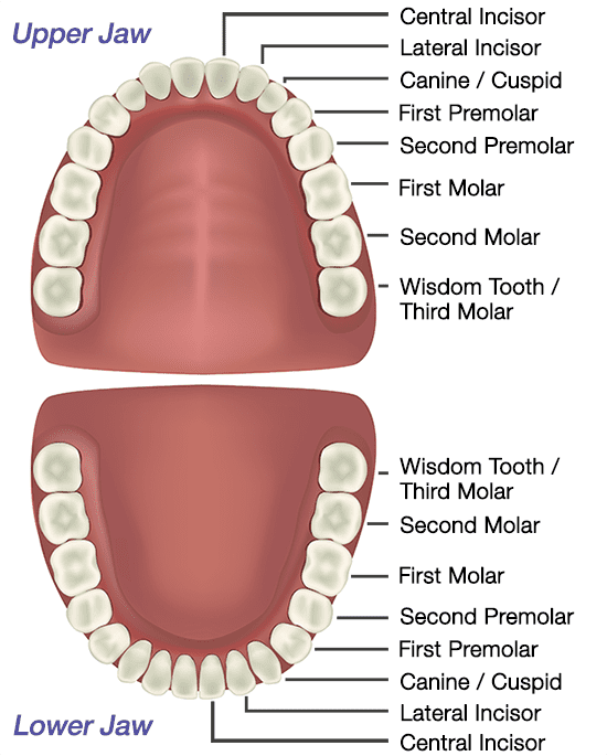
The permanent dentition consists of 32 teeth. This is made up of four incisors, two canines (or cuspids), four premolars (or bicuspids), four molars and two wisdom teeth (also called third molars) in each jaw. If wisdom teeth have been removed there will be 28 teeth.
The incisors are the middlemost four teeth on the upper and lower jaws. They are used for cutting, tearing and holding food. The biting section of an incisor is wide and thin, making a chisel-shaped cutting edge.
The canines (or cuspids, meaning a tooth with a single point) are on either side of the incisors. They are for holding and tearing food.
Premolars (bicuspids) and molars have a series of elevations (points or ‘cusps’) that are used for breaking up particles of food. Each premolar generally has two cusps, hence the name bicuspid. They are used for holding and crushing food.
Molars are the flat teeth at the rear of the mouth. Each molar typically has four or five cusps. They are used exclusively for crushing and grinding.
Wisdom teeth are also called third molars. They erupt from the age of 18 onwards but are often surgically removed.
How many roots does each tooth have?
The number of roots for each type of tooth varies. Typically incisors, canines and premolars will have one root whereas molars will have two or three.
What do my teeth look like?
Our diagrams and images below will show you what the crown and roots of your Incisor, Canine (Cuspid), Premolar (Bicuspid) and Molar teeth look like in each jaw:
Incisor teeth:
You have four incisor teeth (2x central and 2x lateral incisors) in each jaw. They look like this:

Canine teeth:
You have two canine (or cuspid) teeth in each jaw. They look like this:

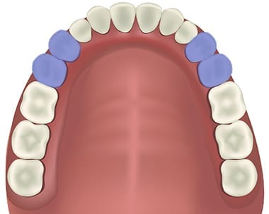
Premolar teeth:
You have four premolar (or bicuspid) teeth in each jaw. They look like this:
Molar teeth:
You have six molars in each jaw. These are made up of four molars (‘first’ and ‘second’ molars), and two wisdom teeth (also called ‘third molars’). They look like this:

What are teeth made of?
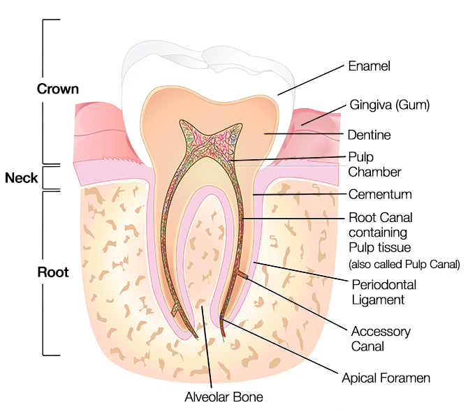
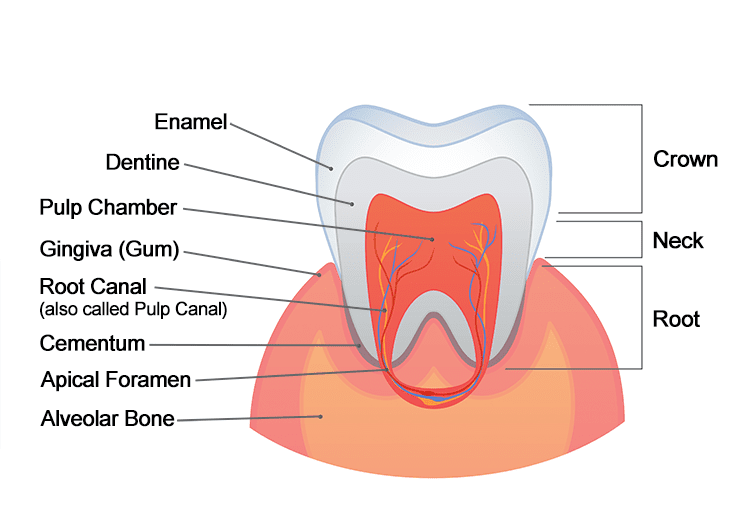
The diagram below shows a cross sectional view of a tooth.
Crown, Root and Neck:
The tooth has two anatomical parts, the crown and the root.
The crown of a tooth is the top part that is exposed and visible above the gum (gingiva). It is covered with enamel, which protects the underlying dentine.
The root of a tooth descends below the gum line, into the upper or lower jawbones, anchoring the tooth in the mouth. Different types of teeth have a different number of roots and root formations. Typically incisors, canines and premolars will have one root whereas molars will have two or three.
The neck is the dividing region of tooth at the gum line, where the crown meets the root.
Enamel:
The crown of each tooth is covered with enamel, which protects the underlying dentine. Enamel is the hardest substance in the human body, even harder than bone. This is because it is the most mineralised substance in the body, made up of crystalline calcium phosphate (Hydroxylapatite). It is as hard as crystal.
Enamel is the only tissue that has no living cells. Because it is not alive, it can’t repair itself from decay or damage.
Gingiva (Gum):
The gingiva is the pink soft tissue that we call our gums. It protects the jaw (alveolar) bone and roots of the teeth, and covers the neck of each tooth.
Dentine:
Dentine forms the major component of each tooth, and extends almost the entire length of the tooth. It is a living tissue, softer than enamel with a structure similar to bone. In contrast to the brittle nature of enamel, dentine is elastic and compressible. It is sensitive, and is protected by enamel on the crown portion and cementum on the roots. It is nourished by the pulp.
Pulp Chamber:
The pulp chamber is the innermost portion of the tooth, lying beneath the dentine and extending from the crown to the tip of the root. The pulp chamber holds the pulp, which is made up of soft tissue. It contains blood vessels to supply blood and nutrients to the tooth to keep it alive, and nerves to enable the tooth to sense temperature. It also contains small lymph vessels carrying white blood cells to the tooth to help fight bacteria.
Cementum:
The cementum is a layer of hard tissue that covers the root of the tooth. It is roughly as hard as bone but considerably softer than enamel. The connective tissues attach to the periodontal ligament, and through this bind the roots of the tooth to the gums and jaw (alveolar) bone.
Root Canal/ Pulp Canal:
The root canal (also called the pulp canal) is the open space inside the root where the pulp extends from the pulp chamber. Blood vessels and nerves from surrounding outside tissue enter the pulp through the root canal.
Periodontal Ligament:
The periodontal ligament is comprised of bundles of connective tissue fibres. One end of each bundle is attached to the cementum covering the root of the tooth. The fibres on the other end anchor the tooth root to the jaw (alveolar) bone and act as shock absorbers, allowing the tooth to withstand the forces of biting and chewing.
Accessory Canal:
Accessory canals are smaller channels that branch off from the main root canal through the dentine to the periodontal ligament. They are usually found near the root end of the tooth (apex). They supply blood vessels and nerves to the pulp.
Apical Foramen:
The apical foramen is the tiny opening at the tip of each root. This is what blood vessels and nerves from surrounding outside tissue pass through to enter the tooth.
Alveolar bone:
The alveolar bone is the jaw bone that surrounds and supports the root of the tooth. It contains the tooth sockets within which the tooth roots are embedded.

Children’s teeth:
Children’s deciduous teeth are also called baby, milk or primary teeth. They are the first set of teeth we receive and will eventually fall out and be replaced with a second set.
When do the first teeth start coming through?
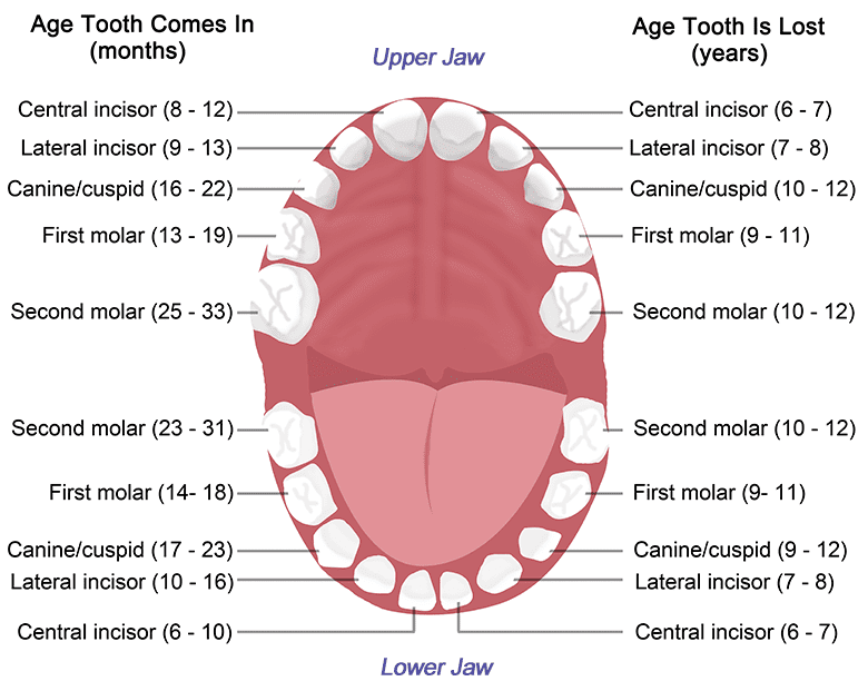
Primary teeth start to form when the baby is in the womb, but start to come through the gums (erupt) when the child is between 6 – 12 months old. Children should have their complete set by 3 years old.
Teeth tend to erupt in parallel, so for example the top molar on the left side should grow in at about the same time as the top molar on the right.
How many teeth do children have, and what are their different types and functions?
The primary set of teeth consists of 20 teeth.
These are made up of: four incisors, two canines and four molars in each jaw.
Incisors bite pieces of food, canines hold and tear food apart and molars grind food.
The diagram below shows where the teeth grow in the mouth:
In adult dentition (the second set of teeth) the 8 primary molars are replaced by the premolar (or bicuspid) teeth. The 12 adult molars erupt (grow up from the gums) behind the primary teeth and do not replace any; giving a total of 32 teeth. The adult dentition is therefore made up of four incisors, two canines, four premolars and six molars in each jaw.
When do children start losing their primary teeth?
Primary teeth are usually replaced by adult teeth between the ages of 6 and 12.
How are primary teeth different from permanent (adult) teeth?
Primary teeth are smaller, have more pointed cusps and are a whiter colour than permanent teeth. They also have thinner enamel and dentine so are more prone to wear, and have relatively large pulp chambers and small delicate roots.
What are my child’s teeth made of?
Crown, Root and Neck:
The tooth has two anatomical parts, the crown and the root.
The crown is the top part that is exposed and visible above the gum (gingiva). It is covered with enamel, which protects the underlying dentine.
The root of a tooth descends below the gum line anchoring the tooth in the mouth.
The neck is the dividing region of tooth at the gum line, where the crown meets the root.
Enamel:
The crown of each tooth is covered with enamel, which protects the underlying dentine. Enamel is the hardest substance in the human body, even harder than bone.
Enamel is the only tissue that has no living cells. Because it is not alive, it can’t repair itself from decay or damage.
Gingiva (Gum):
The gingiva is the pink soft tissue that we call our gums. It protects the jaw (alveolar) bone and roots of the teeth, and covers the neck of each tooth.
Dentine:
Dentine forms the major component of each tooth, and extends almost the entire length of the tooth. It is a living tissue, softer than enamel with a structure similar to bone. It is sensitive, and is protected by enamel on the crown portion and cementum on the roots. It is nourished by the pulp.
Pulp Chamber:
The pulp chamber is the innermost portion of the tooth. The pulp chamber holds the pulp, which is made up of soft tissue. It contains blood vessels to supply blood and nutrients to the tooth to keep it alive, and nerves to enable the tooth to sense temperature.
Cementum:
The cementum is a layer of hard tissue that covers the root of the tooth. It is roughly as hard as bone but considerably softer than enamel. The connective tissues attach to the periodontal ligament, and through this bind the roots of the tooth to the gums and jaw (alveolar) bone.
Root Canal/ Pulp Canal:
The root canal (also called the pulp canal) is the open space inside the root where the pulp extends from the pulp chamber. Blood vessels and nerves from surrounding outside tissue enter the pulp through the root canal.
Periodontal Ligament:
The periodontal ligament is comprised of bundles of connective tissue fibres. One end of each bundle is attached to the cementum covering the root of the tooth. The fibres on the other end anchor the tooth root to the jaw (alveolar) bone and act as shock absorbers, allowing the tooth to withstand the forces of biting and chewing.
Accessory Canal:
Accessory canals are smaller channels that branch off from the main root canal. They are usually found near the root end of the tooth (apex). They supply blood vessels and nerves to the pulp.
Apical Foramen:
The apical foramen is the tiny opening at the tip of each root. This is what blood vessels and nerves from surrounding outside tissue pass through to enter the tooth.
Alveolar bone:
The alveolar bone is the jaw bone that surrounds and supports the root of the tooth. It contains the tooth sockets within which the tooth roots are embedded.
The general information provided by VC Dental is intended as a guide only. It is not to be taken as personal, professional advice. Before making any decision regarding your dental or medical health, it is important to consult with your dentist or medical practitioner.