WO2001096340A1 - Method for inactivation of microorganisms using photosensitizers - Google Patents
Method for inactivation of microorganisms using photosensitizers Download PDFInfo
- Publication number
- WO2001096340A1 WO2001096340A1 PCT/US2001/018752 US0118752W WO0196340A1 WO 2001096340 A1 WO2001096340 A1 WO 2001096340A1 US 0118752 W US0118752 W US 0118752W WO 0196340 A1 WO0196340 A1 WO 0196340A1
- Authority
- WO
- WIPO (PCT)
- Prior art keywords
- fluid
- photosensitizer
- light
- photoradiation
- plasma
- Prior art date
Links
Classifications
-
- A—HUMAN NECESSITIES
- A61—MEDICAL OR VETERINARY SCIENCE; HYGIENE
- A61L—METHODS OR APPARATUS FOR STERILISING MATERIALS OR OBJECTS IN GENERAL; DISINFECTION, STERILISATION OR DEODORISATION OF AIR; CHEMICAL ASPECTS OF BANDAGES, DRESSINGS, ABSORBENT PADS OR SURGICAL ARTICLES; MATERIALS FOR BANDAGES, DRESSINGS, ABSORBENT PADS OR SURGICAL ARTICLES
- A61L2/00—Methods or apparatus for disinfecting or sterilising materials or objects other than foodstuffs or contact lenses; Accessories therefor
- A61L2/0005—Methods or apparatus for disinfecting or sterilising materials or objects other than foodstuffs or contact lenses; Accessories therefor for pharmaceuticals, biologicals or living parts
- A61L2/0011—Methods or apparatus for disinfecting or sterilising materials or objects other than foodstuffs or contact lenses; Accessories therefor for pharmaceuticals, biologicals or living parts using physical methods
- A61L2/0029—Radiation
- A61L2/0076—Radiation using a photocatalyst or photosensitiser
-
- A—HUMAN NECESSITIES
- A61—MEDICAL OR VETERINARY SCIENCE; HYGIENE
- A61L—METHODS OR APPARATUS FOR STERILISING MATERIALS OR OBJECTS IN GENERAL; DISINFECTION, STERILISATION OR DEODORISATION OF AIR; CHEMICAL ASPECTS OF BANDAGES, DRESSINGS, ABSORBENT PADS OR SURGICAL ARTICLES; MATERIALS FOR BANDAGES, DRESSINGS, ABSORBENT PADS OR SURGICAL ARTICLES
- A61L2/00—Methods or apparatus for disinfecting or sterilising materials or objects other than foodstuffs or contact lenses; Accessories therefor
- A61L2/0005—Methods or apparatus for disinfecting or sterilising materials or objects other than foodstuffs or contact lenses; Accessories therefor for pharmaceuticals, biologicals or living parts
- A61L2/0011—Methods or apparatus for disinfecting or sterilising materials or objects other than foodstuffs or contact lenses; Accessories therefor for pharmaceuticals, biologicals or living parts using physical methods
-
- A—HUMAN NECESSITIES
- A61—MEDICAL OR VETERINARY SCIENCE; HYGIENE
- A61L—METHODS OR APPARATUS FOR STERILISING MATERIALS OR OBJECTS IN GENERAL; DISINFECTION, STERILISATION OR DEODORISATION OF AIR; CHEMICAL ASPECTS OF BANDAGES, DRESSINGS, ABSORBENT PADS OR SURGICAL ARTICLES; MATERIALS FOR BANDAGES, DRESSINGS, ABSORBENT PADS OR SURGICAL ARTICLES
- A61L2/00—Methods or apparatus for disinfecting or sterilising materials or objects other than foodstuffs or contact lenses; Accessories therefor
- A61L2/0005—Methods or apparatus for disinfecting or sterilising materials or objects other than foodstuffs or contact lenses; Accessories therefor for pharmaceuticals, biologicals or living parts
- A61L2/0082—Methods or apparatus for disinfecting or sterilising materials or objects other than foodstuffs or contact lenses; Accessories therefor for pharmaceuticals, biologicals or living parts using chemical substances
- A61L2/0088—Liquid substances
-
- A—HUMAN NECESSITIES
- A61—MEDICAL OR VETERINARY SCIENCE; HYGIENE
- A61L—METHODS OR APPARATUS FOR STERILISING MATERIALS OR OBJECTS IN GENERAL; DISINFECTION, STERILISATION OR DEODORISATION OF AIR; CHEMICAL ASPECTS OF BANDAGES, DRESSINGS, ABSORBENT PADS OR SURGICAL ARTICLES; MATERIALS FOR BANDAGES, DRESSINGS, ABSORBENT PADS OR SURGICAL ARTICLES
- A61L2/00—Methods or apparatus for disinfecting or sterilising materials or objects other than foodstuffs or contact lenses; Accessories therefor
- A61L2/02—Methods or apparatus for disinfecting or sterilising materials or objects other than foodstuffs or contact lenses; Accessories therefor using physical phenomena
- A61L2/08—Radiation
- A61L2/10—Ultra-violet radiation
-
- A—HUMAN NECESSITIES
- A61—MEDICAL OR VETERINARY SCIENCE; HYGIENE
- A61L—METHODS OR APPARATUS FOR STERILISING MATERIALS OR OBJECTS IN GENERAL; DISINFECTION, STERILISATION OR DEODORISATION OF AIR; CHEMICAL ASPECTS OF BANDAGES, DRESSINGS, ABSORBENT PADS OR SURGICAL ARTICLES; MATERIALS FOR BANDAGES, DRESSINGS, ABSORBENT PADS OR SURGICAL ARTICLES
- A61L2202/00—Aspects relating to methods or apparatus for disinfecting or sterilising materials or objects
- A61L2202/20—Targets to be treated
- A61L2202/22—Blood or products thereof
Definitions
- hepatitis and other viruses and bacteria presents a serious health hazard for those who must receive transfusions of whole blood or administration of various blood components such as platelets, red cells, blood plasma, Factor NIII, plasminogen, fibronectin, anti-thrombin III, cryoprecipitate, human plasma protein fraction, albumin, immune serum globulin, prothrombin complex, plasma growth hormones, and other components isolated from blood.
- various blood components such as platelets, red cells, blood plasma, Factor NIII, plasminogen, fibronectin, anti-thrombin III, cryoprecipitate, human plasma protein fraction, albumin, immune serum globulin, prothrombin complex, plasma growth hormones, and other components isolated from blood.
- Blood screening procedures may miss contaminants, and sterilization procedures which do not damage cellular blood components but effectively inactivate all infectious viruses and other microorganisms have not heretofore been available.
- Solvent detergent methods of blood component decontamination work by dissolving phospholipid membranes surrounding viruses such as HIN, and do not damage protein components of blood; however, if blood cells are present, such methods cannot be used because of damage to cell membranes.
- photosensitizers compounds which absorb light of a defined wavelength and transfer the absorbed energy to an energy acceptor
- European Patent application 196,515 published October 8, 1986, suggests the use of non-endogenous photosensitizers such as porphyrins,
- S5 psoralens S5 psoralens, acridine, toluidines, flavine (acriflavine hydrochloride), phenothiazine derivatives, and dyes such as neutral red and methylene blue, as blood additives.
- Protoporphyrin which occurs naturally within the body, can be metabolized to form a photosensitizer; however, its usefulness is limited in that it degrades desired biological activities of proteins.
- Chlorpromazine is also exemplified as one such photosensitizer; however its usefulness is limited by the fact that it should be removed from any fluid administered to a patient after the decontamination procedure because it has a sedative effect.
- Patent 4,727,027 issued February 23, 1988 to Wiesehahn, G.P., et al. discloses the use of furocoumarins including psoralen and derivatives for decontamination of blood and blood products, but teaches that steps must be taken to reduce the availability of dissolved oxygen and other reactive species in order to inhibit denaturation of biologically active proteins.
- Photoinactivation of viral and bacterial blood contaminants using halogenated coumarins is described in U.S. Patent 5,516,629 issued May 14, 1996 to Park, et al.
- Patent 5,232,844 issued August 3, 1993 to Horowitz, et al. also disclose the need for the use of "quenchers” in combination with photosensitizers which attack lipid membranes, and U.S. Patent 5,360,734 issued November 1, 1994 to Chapman et al. also addresses the problem of prevention of damage to other blood components.
- Riboflavin (7,8-dimethyl-lO-ribityl isoalloxazine) has been reported to attack nucleic acids. Photoalteration of nucleic acid in the presence of riboflavin is discussed in Tsugita, A, et al. (1965), "Photosensitized inactivation of ribonucleic acids in the presence of riboflavin,"
- U.S. Patent No. 5,290,221 issued March 1, 1994 to Wolfe, Jr., et al.
- U.S. Patent No. 5,536,238 issued July 16, 1996 to Bischof.
- U.S. Patent No. 5,290,221 discloses the irradiation of fluid in a relatively narrow, arcuate gap.
- U.S. Patent 5,536,238 discloses devices utilizing optical fibers extending into a filtration medium. Both patents recommend as photosensitizers benzoporphryin derivatives which have an affinity for cell walls.
- U.S. Patent No. 5,527,704 issued June 18, 1996 to Wolf, Jr., et al. discusses an apparatus to inactivate viruses contained in a body fluid in a container using methylene blue as a photosensitizer. The body fluid is maintained in a static state within the container during irradiation.
- U.S. Patent No. 5,868,695 issued February 9, 1999 to Wolf, Jr. et al. discloses a system where blood containing a photoactive material is directed in a predetermined flow path such as a serpentine in a narrow gap in a treatment chamber.
- PCT published application No. WO 96/06647 discloses irradiating a product in an array of light emitting diodes surrounded by a fluid used to prevent overheating of the diodes.
- U. S. Patents 5,360,734 issued November 1, 1994 to Chapman et al. and 5,597,722 issued January 28, 1997 to Chapman et al. discuss treating a blood component containing red blood cells and plasma proteins with a photoactive agent such as pyrrolic macrocycles, psoralens or methylene blue after removing a portion of the plasma proteins.
- the treated blood component is required to be prevented from contacting plasma proteins for a period of time (three to eighteen hours) after treatment to prevent bonding of the treated cells with IgG proteins in tne plasma.
- 1 ne presence ot IgG has various negative effects, including interference with commonly used serological and diagnostic testing procedures.
- Methods and apparatuses are provided for treating a fluid to inactivate at least some of the microorganisms and/or white cells that may be present therein or thereon, said fluid containing one or more components selected from the group consisting of protein, (e.g. biologically active protein such as a therapeutic protein), blood and blood constituents.
- protein e.g. biologically active protein such as a therapeutic protein
- blood and blood constituents e.g. blood and blood constituents.
- the adjusting step may be any method useful to adjust the amount of plasma present in a sample, or any method useful to reduce the amount of a particular component of plasma in a sample.
- One method suitable for the adjusting step is diluting the fluid with a diluting solution. This will reduce the level of all plasma constituents.
- the diluting solution used to bring the level of plasma to a desired value may be one of many different solutions, including saline; a buffer, which may comprise a variety of different substances; a solution containing glucose, phosphate or both, which may or may not act as a buffer; a solution containing nutrients for a component of the sample; a cryopreservative; an anticoagulant; a cell storage solution known to the art or developed to provide cells with suitable additives to enable them to be stored or infused; or other suitable solution.
- the solution should not substantially interfere with the inactivation of microorganisms or substantially destroy the biological activity of the fluid.
- the solution does not "substantially interfere.”
- the solution does not "substantially destroy" the biological activity.
- Selected proteins in plasma may be reduced selectively by proper selection of reagents and filtration.
- reagents and methods of filtering are known in the art.
- One such filter is a hollow fiber filter.
- the adjusting step may also be carried out using a mechanical means such as by centrifuging the fluid to separate the components, collecting the desired component or components and resuspending the desired component or components in a suitable solution. The process may be carried out more than once, to achieve the desired level of plasma adjustment.
- the process may also be carried out using gravity to separate the components.
- the adjusting step may also comprise washing the fluid one or more times, as known in the art. Washing is generally adding a solution to reduce the level of plasma in extracellular space between the cells.
- the fluid may be washed with a detergent solution or saline, for example.
- the amount of one component of plasma may be reduced selectively.
- the amount of bilirubin in a sample may be reduced by filtration through resin cartridges or washing or pre-irradiation with 447 nm light to break down the bilirubin, or any other method known to the art.
- the amount of the total plasma in the sample may be reduced. Both of these embodiments are intended to be encompassed by terms such as "reducing the percentage of plasma" and the like.
- the mixing step may occur before, after or simultaneously with the adjusting step.
- the photosensitizer may be a photo-activatable compound whose photolytic products
- the most preferred photosensitizer is 7,8-dimethyl- 10-ribityl isoalloxazine.
- the percentage of plasma in the fluid in the methods of the invention may be adjusted to any desired level that provides the effect of measurably improving the inactivation of microorganisms as compared to a fluid of the same composition if the percentage of plasma had not been reduced.
- Improving the inactivation of microorganisms means the light dose required for a desired level of inactivation is lower, the wavelength range of light required to inactivate a desired number of microorganism is narrower, the level of photosensitizer required is lower, the time required for a desired level of microorganism inactivation is shorter than required in a fluid of the same composition if the percentage of plasma had not been reduced, or a combination of these effects.
- Photoradiation may comprise light in the visible spectrum, the ultraviolet spectrum, or light in both the visible and ultraviolet spectra. Any suitable wavelength or wavelengths of light may be used in any proportion and energy that produces the desired level of inactivation of microorganisms. For example, about half the light may be in the visible spectrum and about half the light may be in the ultraviolet spectrum. Alternatively, about one third of light may be in one spectral range and the other two thirds of light in the other spectral range. In one embodiment of the invention, two light sources (or two arrays of light sources) are used, to provide two wavelengths of light. As used herein, "wavelength" does not necessarily mean one discrete wavelength. Wavelength may comprise a range of about ⁇ 100 nm centered around one wavelength. Preferably, if ultraviolet light is used, the amount of ultraviolet light is kept to a level that minimizes damage to desired fluid components. Generally, this is provided by using 50% or less ultraviolet light relative to the total light energy delivered.
- the fluid containing the photosensitizer is exposed to photoradiation of the appropriate wavelength to activate the photosensitizer, using an amount of photoradiation sufficient to activate the photosensitizer as described herein, but less than that which would cause non-specific damage to the biological components or substantially interfere with biological activity of other proteins present in the fluid.
- the wavelength used will depend on the photosensitizer selected and composition of the fluid, as is known to the art or readily determinable without undue experimentation following the teachings disclosed herein.
- Nonspecific damage is damage that damages all components.
- the photoradiation in both the ultraviolet and visible spectra may be supplied concurrently or sequentially, with the visible portion preferably being supplied first.
- the photoradiation source may be a simple lamp or may consist of multiple lamps radiating at differing wavelengths.
- the photoradiation source should be capable of delivering a sufficient amount of light to activate the photosensitizer, preferably from about 100 to at least about 200 J/cm 2 .
- an apparatus for inactivating microorganisms which may be present in a fluid with an endogenous or endogenously-based derivative photosensitizer, comprising:
- the means for maintaining the fluid and an effective amount of an endogenous or endogenously-based derivative photosensitizer in the light path may comprise a support surface substantially parallel to said source of light; a cuvette and flow-through system; or other means known in the art.
- a system for treating a fluid to inactivate microorganisms which may be present therein with an endogenous or endogenously based derivative photosensitizer comprising:
- a container comprising said fluid having a desired level of plasma, at least an effective amount of an endogenous photosensitizer or endogenously-based derivative photosensitizer, and optionally one or more additives, said container having a photopermeable surface sufficient to allow exposure of the fluid therein to an amount of photoradiation sufficient to activate the photosensitizer;
- At least one photoradiation source in light communication with said container, said source capable of generating a suitable wavelength and intensity to activate the endogenous photosensitizer or endogenously-based derivative photosensitizer whereby microorganisms present are inactivated.
- Also provided is a system for inactivation of microorganisms in a fluid containing such microorganisms comprising:
- At least one photoradiation source for providing sufficient photoradiation to the fluid in said container of a type and amount selected to activate the photosensitizer.
- a system for treating a fluid to inactivate microorganisms comprising:
- At least one photoradiation source in light communication with said container, said source capable of providing sufficient photoradiation to the fluid in said container of a type and amount selected to activate the photosensitizer whereby microorganisms are inactivated.
- the photopermeable container may be a transparent plastic bag, a transparent plastic container with rigid walls, or other containers as known to the art.
- the agitation may be provided by a shaker table, or other means for agitating known to the art.
- a light source that emits light of a suitable wavelength and intensity to inactivate microorganisms which may be present in said fluid.
- the term "inactivation of a microorganism” means totally or partially preventing the microorganism from replicating, either by killing the microorganism or otherwise interfering with its ability to reproduce.
- Microorganisms include viruses (both extracellular and intracellular), bacteria, bacteriophages, fungi, blood-transmitted parasites, and protozoa.
- Exemplary viruses include acquired immunodeficiency (HIN) virus, hepatitis A, B and C viruses, sinbis virus, cytomegalovirus, vesicular stomatitis virus, herpes simplex viruses, e.g. types I and II, human T-lymphotropic retroviruses, HTLN-III, lymphadenopathy virus LAN/ID AN, parvovirus, transfusion-transmitted (TT) virus, Epstein-Barr virus, and others known to the art.
- HIN acquired immunodeficiency
- hepatitis A, B and C viruses sinbis virus
- cytomegalovirus vesicular stomatitis virus
- herpes simplex viruses e.g. types I and II, human T-lymphotropic retroviruses, HTLN-III, lympha
- Bacteriophages include ⁇ X174, ⁇ 6, ⁇ , R17, T 4 , and T 2 .
- Exemplary bacteria include P. aeruginosa, S. aureus, S. epidermis, L. monocytogenes, E. coli, K. pneumonia and S. marcescens.
- Inactivation of white blood cells may be desirable when suppression of immune or autoimmune response is desired, e.g., in processes involving transfusion of red cells, platelets or plasma when donor white blood cells may be present.
- the process disclosed may be used to inactivate white blood cells.
- Materials which may be treated by the methods of this invention include any materials which are adequately permeable to photoradiation to provide sufficient light to achieve microorganism inactivation, or which can be suspended or dissolved in fluids which have such permeability to photoradiation.
- materials are whole blood and aqueous compositions containing biologically active proteins derived from blood or blood constituents. Packed red cells, platelets and plasma (fresh or fresh frozen plasma) are exemplary of such blood constituents.
- therapeutic protein compositions containing proteins derived from blood such as fluids containing biologically active protein useful in the treatment of medical disorders, e.g.
- factor VIII Von Willebrand factor
- factor IX factor X
- factor XI factor XI
- Hageman factor prothrombin
- anti-thrombin III fibronectin
- plasminogen plasma protein fraction
- immune serum globulin modified immune globulin
- albumin plasma growth hormone
- somatomedin plasminogen streptokinase complex
- ceruloplasmin transferrin
- haptoglobin antitrypsin and prekallikrein
- prekallikrein may be treated by the decontamination methods of this invention.
- the activity of a biologically-active protein in said fluid is at a biologically-active level after said exposing step.
- a therapeutic protein present in said fluid remains able to perform a therapeutic function after the exposing step.
- Other fluids which could benefit from the treatment of this invention are peritoneal solutions used for peritoneal dialysis which are sometimes contaminated during connection, leading to peritoneal infections.
- biologically active means capable of effecting a change in a living organism or component thereof.
- biologically active with respect to “biologically active protein” as referred to herein does not refer to proteins which are part of the microorganisms being inactivated.
- non-toxic with respect to the photosensitizers means low or no toxicity to humans and other mammals, and does not mean non-toxic to the microorganisms being inactivated.
- Substantial destruction" of biological activity means at least as much destruction as is caused by porphyrin and porphyrin derivatives, metabolites and precursors which are known to have a damaging effect on biologically active proteins and cells of humans and mammals.
- substantially non-toxic means less toxic than porphyrin, porphyrin derivatives, metabolites and precursors that are known for blood sterilization.
- blood product includes blood constituents and therapeutic protein compositions containing proteins derived from blood as defined above. Fluids containing biologically active proteins other than those derived from blood may also be treated by the methods of this invention.
- Decontamination methods of this invention using endogenous photosensitizers and endogenously-based photosensitizer derivatives do not substantially destroy the biological activity of fluid components other than microorganisms. As much biological activity of these components as possible is retained, although in certain instances, when the methods are optimized, some loss of biological activity, e.g., denaturization of protein components, must be balanced against effective decontamination of the fluid. So long as fluid components retain sufficient biological activity to be useful for their intended or natural purposes, their biological activities are not considered to be “substantially destroyed.” Photosensitizers are known to be useful for inactivating microorganisms.
- a "photosensitizer” is defined as any compound which absorbs radiation of one or more defined wavelengths and subsequently utilizes the absorbed energy to carry out a chemical process.
- photosensitizers include porphyrins, psoralens, dyes such as neutral red, methylene blue, acridine, toluidines, flavine (acriflavine hydrochloride) and phenothiazine derivatives, coumarins, quinolones, quinones, and anthroquinones.
- Photosensitizers of this invention may include compounds which preferentially adsorb to nucleic acids, thus focusing their photodynamic effect upon microorganisms and viruses with little or no effect upon accompanying cells or proteins.
- photosensitizers are also useful in this invention, such as those using singlet oxygen-dependent mechanisms. Most preferred are endogenous photosensitizers.
- endogenous means naturally found in a human or mammalian body, either as a result of synthesis by the body or because of ingestion as an essential foodstuff (e.g. vitamins) or formation of metabolites and/or byproducts in vivo.
- endogenous photosensitizers are alloxazines such as 7,8-dimethyl-10-ribityl isoalloxazine (riboflavin), 7,8,10-trimethylisoalloxazine (lumiflavin), 7,8-dimethylalloxazine (lumichrome), isoalloxazine-adenine dinucleotide (flavine adenine dinucleotide [FAD]), alloxazine mononucleotide (also known as flavine mononucleotide [FMN] and riboflavine-5- phosphate), vitamin Ks, vitamin L, their metabolites and precursors, and napththoquinones, naphthalenes, naphthols and their derivatives having planar molecular conformations.
- alloxazines such as 7,8-dimethyl-10-ribityl isoalloxazine (riboflavin), 7,8,10-trimethylisoalloxazine (lumif
- alloxazine includes isoalloxazines.
- Endogenously-based derivative photosensitizers include synthetically derived analogs and homologs of endogenous photosensitizers which may have or lack lower (1-5) alkyl or halogen substituents of the photosensitizers from which they are derived, and which preserve the function and substantial non-toxicity thereof.
- endogenous photosensitizers particularly when such photosensitizers are not inherently toxic or do not yield toxic photoproducts after photoradiation, no removal or purification step is required after decontamination, and treated product can be directly returned to a patient's body or administered to a patient in need of its therapeutic effect.
- Non-endogenous photosensitizers based on endogenous structures such as those described in U.S. Patent Application 09/420,652 are also included. These non-endogenous photosensitizers and endogenously-based derivative photosensitizers are referred to herein as endogenously-based derivative photosensitizers.
- binder means dried medium, including powder or pill.
- a dried medium of a substance is described herein, it is also intended that a solution or suspension of the powder in a suitable solvent may be used, and vice versa.
- the method of this invention requires mixing the photosensitizer with the material to be decontaminated. "Adding” is intended to include mixing the fluid with the photosensitizer. Mixing may be done by simply adding the photosensitizer or a solution containing the photosensitizer to a fluid to be decontaminated.
- the material to be decontaminated to which photosensitizer has been added is flowed past a photoradiation source, and the flow of the material generally provides sufficient turbulence to distribute the photosensitizer throughout the fluid to be decontaminated.
- the fluid and photosensitizer are placed in a photopermeable container and irradiated in batch mode, preferably while agitating the container to fully distribute the photosensitizer and expose all the fluid to the radiation.
- the amount of photosensitizer to be mixed with the fluid will be an amount sufficient to adequately inactivate microorganisms therein, but less than a toxic (to humans or other mammals) or insoluble amount. Excess photosensitizer may be used as long as the concentration is not so high that the photosensitizer prevents light from passing to the desired depth at a useful intensity. As taught herein, optimal concentrations for desired photosensitizers may be readily determined by those skilled in the art without undue experimentation. Preferably the photosensitizer is used in a concentration of at least about 1 micromolar. The optimum concentration of photosensitizer will vary depending on the blood component being treated and the level to which plasma is removed.
- red blood cells are being treated, a higher concentration of photosensitizer is desired than if plasma or platelets are being treated.
- a useful concentration of riboflavin is about 1-200 micromolar, and a preferred concentration of riboflavin is about 50 to 150 micromolar when the plasma content is about 0 to 5% of the total volume of the solution.
- a useful concentration of riboflavin is about 1-100 micromolar, and a preferred concentration of riboflavin is about 10 to 30 micromolar when the plasma content is about 10 - 90% of the total volume of the solution.
- the activated photosensitizer is capable of inactivating the microorganisms present, such as by interfering to prevent their replication. Specificity of action of the photosensitizer is conferred by the close proximity of the photosensitizer to the nucleic acid of the microorganism and this may result from binding of the photosensitizer to the nucleic acid.
- "Nucleic acid” includes ribonucleic acid (RNA) and deoxyribonucleic acid (DNA). Other photosensitizers may act by binding to cell membranes or by other mechanisms.
- the photosensitizer may also be targeted to the microorganism to be inactivated by covalently coupling to an antibody, preferably a specific monoclonal antibody to the microorganism.
- the fluid containing the photosensitizer may be flowed into a photopermeable container for irradiation.
- container refers to a closed or open space, which may be made of rigid or flexible material, e.g., may be a bag or box or trough. It may be closed or open at the top and may have openings at both ends, e.g., may be a tube or tubing, to allow for flow-through of fluid therein.
- a cuvette has been used to exemplify one embodiment of the invention involving a flow-through system. Collection bags, such as those used with the TrimaTM SpectraTM and apheresis systems of GAMBRO, Inc., have been used to exemplify another embodiment involving batch- wise treatment of the fluid.
- photopermeable means the material of the container is adequately transparent to photoradiation of the proper wavelength for activating the photosensitizer.
- the container has a depth (dimension measured in the direction of the radiation from the photoradiation source) sufficient to allow photoradiation to adequately penetrate the container to contact photosensitizer molecules at all distances from the light source and ensure inactivation of microorganisms in the fluid to be decontaminated, and a length (dimension in the direction of fluid flow) sufficient to ensure a sufficient exposure time of the fluid to the photoradiation.
- the fluid to be treated is placed in a photopermeable container which is agitated and exposed to photoradiation for a time sufficient to substantially inactivate the microorganisms.
- the photopermeable container is preferably a blood bag made of transparent or semitransparent plastic, and the agitating means is preferably a shaker table.
- the photopermeable container may be any other container, such as a rigid plastic container.
- the photosensitizer may be added to the container in powdered or liquid form and the container agitated to mix the photosensitizer with the fluid and to adequately expose all the fluid to the photoradiation to ensure inactivation of microorganisms.
- Photosensitizer may be added to or flowed into the photopermeable container separately from the fluid being treated or may be added to the fluid prior to placing the fluid in the container.
- photosensitizer is added to anticoagulant ana tne mixture of photosensitizer and anticoagulant are added to the fluid.
- the blood or blood product may be delivered to a patient, concentrated, or infused directly.
- Enhancers may also be added to the fluid to make the process more efficient and selective.
- enhancers include antioxidants or other agents to prevent damage to desired fluid components or to improve the rate of inactivation of microorganisms and are exemplified by adenine, histidine, cysteine, tyrosine, tryptophan, ascorbate, N-acetyl-L- cysteine, propyl gallate, glutathione, mercaptopropionylglycine, dithiothreotol, nicotinamide, BHT, BHA, lysine, serine, methionine, glucose, mannitol, trolox, glycerol, and mixtures thereof.
- These enhancers may be added in dried medium, including powder or pill form or in the form of liquids.
- This invention also comprises fluids comprising biologically active protein, blood or blood constituents and also containing endogenous photosensitizer, endogenously-based derivative photosensitizer, or photoproduct thereof made by the inactivation methods described herein.
- the fluid may also contain inactivated microorganisms.
- this method is useful for treating other fluids including fluids which are meant for nourishment of humans or animals such as water, fruit, juices, milk, broths, soups and the like.
- the method is also useful for treating peritoneal or parenteral solutions.
- the photoradiation source may be connected to the photopermeable container for the fluid by means of a light guide such as a light channel or fiber optic tube which prevents scattering of the light between the source and the container for the fluid, and more importantly, prevents substantial heating of the fluid within the container.
- a light guide such as a light channel or fiber optic tube which prevents scattering of the light between the source and the container for the fluid, and more importantly, prevents substantial heating of the fluid within the container.
- Direct exposure to the light source may raise temperatures as much as 10 to 15 C, especially when the amount of fluid exposed to the light is small, which can cause denaturization of blood components.
- Use of the light guide keeps any heating to less than about 2 °C.
- the method may also include the use of temperature sensors and cooling mechanisms where necessary to keep the temperature below temperatures at which desired proteins in the fluid are damaged. Cooling mechanisms include flow of air or fluid, as well as otlier mechanisms known to the art.
- the temperature is kept between about 0°C and about 45°C, more
- any means for adding the photosensitizer to the fluid to be decontaminated and for placing the fluid in the photopermeable container known to the art may be used, such means typically including flow conduits, ports, reservoirs, sterile docking, valves, and the like.
- the system may include means such as pumps or adjustable valves for controlling the flow of the photosensitizer into the fluid to be decontaminated so that its concentration may be controlled at effective levels as described herein.
- photosensitizer is mixed with the anticoagulant feed to a blood apheresis system.
- the pH of the solution is preferably kept low enough, as is known to the art, to prevent detachment of the sugar moiety.
- the photosensitizer is added to the fluid to be decontaminated in a pre-mixed aqueous solution, e.g., in water or storage buffer solution.
- the photosensitizer and any optional desired additives may be placed in a container as dried medium, including powder or pill form, or as a solution.
- Desired additives include nutrients or other materials such as acetate, glucose, dextrose, citrate, pyruvate, which allow the components to retain biological activity or improve the storage lifetime. It may be desirable for platelets to be provided nutrients when the plasma concentration is less than about 20% of the total volume of the sample in order for the platelets to remain active. Desired additives and the photosensitizer may be sterilized as powders. In one embodiment, the powders desired are placed in the container prior to introduction of the fluid.
- the volume and composition of the solution(s) may produce the desired percentage of plasma in the sample, without further additions of solution, or the percentage of plasma may be adjusted before, during or after placing said fluid in said container. Adjustment of the percentage of plasma after placing the fluid in the container may occur by the introduction of a suitable solution after the fluid is in the container. Adjustment of the percentage of plasma may occur during introduction of the fluid in a container by the introduction of a suitable solution as the fluid is being placed in the container.
- the photopermeable container for the flow-through system may be a transparent cuvette made of polycarbonate, glass, quartz, polystyrene, polyvinyl chloride, polyolefm, or other transparent material.
- the cuvette may be enclosed in a radiation chamber having mirrored walls.
- a photoradiation enhancer such as a second photoradiation source or reflective surface may be placed adjacent to the cuvette to increase t ⁇ e amount of photoradiation contacting the fluid within the cuvette.
- the system preferably includes a pump for adjusting the flow rate of the fluid to desired levels to ensure substantial decontamination as described above.
- the cuvette has a length, coordinated with the flow rate therethrough, sufficient to expose fluid therein to sufficient photoradiation to effect substantial decontamination thereof.
- the cuvette is spaced apart from the light source a sufficient distance that heating of the fluid in the cuvette does not occur, and light is transmitted from the light source to the cuvette by means of a light guide.
- the fluid is placed in a photopermeable container such as a blood bag, e.g. used with the apheresis system described in U.S. Patent No. 5,653,887, and agitated while exposing to photoradiation.
- a photopermeable container such as a blood bag
- Suitable bags include collection bags as described herein. Collection bags used in the SpectraTM system or TrimaTM apheresis system of
- Shaker tables are known to the art, e.g. as described in U.S. Patent 4,880,788.
- the bag is equipped with at least one port or opening for adding fluid thereto.
- the photosensitizer preferably 7,8-dimethyl-10-ribityl- isoalloxazine
- the bag is then placed on a shaker table and agitated under photoradiation until substantially all the fluid has been exposed to the photoradiation.
- the bag may be prepackaged with the powdered photosensitizer contained therein.
- the fluid to be decontaminated may then be added through the appropriate port.
- Decontamination systems as described above may be designed as stand-alone units or may be easily incorporated into existing apparatuses known to the art for separating or treating blood being withdrawn from or administered to a patient.
- blood- handling apparatuses include the GAMBRO SpectraTM or TRIMA ® apheresis systems, available from GAMBRO Inc., Lakewood, CO, or the apparatuses described in U.S. Patent 5,653,887 and U.S. Serial No. 08/924,519 filed September 5, 1997 (PCT Publication No. WO
- the decontamination system may be inserted just downstream of the point where blood is withdrawn from a patient or donor, just prior to insertion of blood product into a patient, or at any point before or after separation of blood constituents.
- the plasma may be adjusted at any point before fluid is exposed to irradiation.
- the photosensitizer is added to blood components along with anticoagulant in a preferred embodiment, and separate irradiation sources and cuvettes are placed downstream from collection points for platelets, for plasma and for red blood cells.
- decontamination systems of this invention may be used to process previously collected and stored blood products.
- the fluid may be thinned, exposed to higher energies of radiation for longer periods, agitated for longer periods or presented to photoradiation in shallower containers or conduits than necessary for use with other blood components.
- the endogenous photosensitizers and endogenously-based derivative photosensitizers disclosed herein can be used in pre-existing blood component decontamination systems as well as in the decontamination system disclosed herein.
- the endogenous photosensitizers and endogenously-based derivative photosensitizers of this invention can be used in the decontamination systems described in U.S. Patent Nos. 5,290,221, 5,536,238, 5,290,221 and 5,536,238.
- Platelet additive solutions comprising endogenous photosensitizers and endogenously-based derivative photosensitizers as described above are also provided herein.
- Platelet additive solutions known to the art may be used for this purpose and include those disclosed in U.S. Patent Nos. 5,908,742; 5,482,828; 5,569,579; 5,236,716; 5,089,146; and 5,459,030.
- Such platelet additive solutions may contain physiological saline solution, buffer, preferably sodium phosphate, and other components including magnesium chloride and sodium gluconate.
- the pH of such solutions is preferably between about 7.0 and 7.4. These solutions are useful as carriers for platelet concentrates to allow maintenance of cell quality and metabolism during storage, reduce plasma content and extend storage life.
- the photosensitizer may be present in such solutions at any desired concentration from about 1 ⁇ M to the solubility of the photosensitizer in the solution, and preferably between about 10 ⁇ M and about 100 ⁇ M, more preferably about 10 ⁇ M.
- the platelet additive solution also comprises enhancers as described above.
- a preferred platelet additive solution comprises sodium acetate, sodium chloride, sodium gluconate, 1.5 micromolar magnesium chloride, 1 micromolar sodium phosphate 14 ⁇ M 7,8-dimethyl-10- ribityl-isoalloxazine and preferably also 6 micromolar ascorbate.
- platelet additive solutions may be added in a desired volume to a fluid so that the level of plasma is at a desired level before irradiation, or platelet additive solutions may oe use ⁇ to resuspend a pellet.
- Other uses for platelet additive solutions are known to the art.
- a photoradiation enhancer such as a reflective surface may also be provided in any method or apparatus of the invention.
- the light may be guided to impinge on the fluid in any desired manner, including positioning the fluid in the light path of the light source, using a light guide, or other methods.
- the apparatuses of the invention may also comprise components such as a temperature monitor, temperature controller, means for flowing said fluid into and out of said container, means for agitating said fluid in said container, and other desired components to control various aspects of the system.
- the temperature controller may be a fan directed toward the light source, directed on the fluid, or both. One or more temperature controllers may be used to cool different components to different levels.
- Figure 1 depicts the riboflavin absorbance spectrum.
- Figure 2 depicts a correlation of light absorbance and hematocrit observed and predicted for red blood cells, and predicted for platelets.
- Figure 3 depicts photodecomposition over time of riboflavin in anticoagulant Acid Citrate Dextrose (ACD) solution.
- the solid line with circles indicates percent of initial riboflavin remaining at 373 nm.
- the dotted line with squares indicates percent of initial riboflavin remaining at 447 nm.
- Figure 4 depicts the transmission profile of various plastic cuvettes as a function of wavelength.
- the solid line represent a 3.2 mm acrylic cuvette.
- the dotted line ( ) represents a 3.2 mm UV acrylic cuvette.
- the dashed line ( ) represents a 3.2 mm polystyrene (PS) cuvette, and the crossed line indicates a 3.2 mm polycarbonate (PC) cuvette.
- PS polystyrene
- PC polycarbonate
- Figure 5 depicts the light flux required in mW per cm 2 as a function of flow rate, i.e. the flux required to deliver one joule/cm 2 to a sample in the cuvette.
- Figure 6 depicts a blood separation apparatus incorporating the photoradiation device of this invention.
- Figure 7 depicts the decontamination assembly of this invention.
- Figure 8 depicts inactivation of bacteria in platelet preparations using vitamin K5 as the photosensitizer as a function of energy of irradiation.
- Figure 9 depicts inactivation of bacteria as a function of platelet preparation and energy of irradiation, using 90% platelets and 10% platelet additive solution (90:10) and 30% platelets with 70% additive solution (30:70).
- Figure 10 shows the effect on inactivation of virus, bacteriophage and bacteria of adding antioxidants to platelet concentrate.
- Figure 11 shows the inactivation curve for Herpes Simplex type II virus as a function of concentration of photosensitizer at an energy of irradiation of 20J/cm 2 using half ultraviolet and half visible light.
- Figure 12 shows inactivation of S. epidermidis at varying concentrations of photosensitizer and energies of irradiation.
- Figure 13 shows inactivation of ⁇ X174 at varying concentrations of photosensitizer and energies of irradiation.
- Figure 14 shows inactivation of S. aureus and ⁇ X174 at varying energies of irradiation using a 50:50 mixture of ultraviolet and visible light.
- Figure 15 shows inactivation of S. epidermidis and HSV-II at varying energies of irradiation using a 50:50 mixture of ultraviolet and visible light.
- Figure 16 shows inactivation of HSV2 virus in blood bags agitated and irradiated at varying energy levels.
- Figure 17 compares inactivation results for vaccinia virus in various fluids using ultraviolet light alone or 50:50 visible and ultraviolet light.
- Figure 18 compares inactivation results with and without sensitizer of vaccinia virus at varying irradiation times.
- Figure 19 compares inactivation of extracellular HIV-1 a ⁇ ' 5 * ahd"50 M o' ⁇ photosensitizer and varying irradiation energies.
- Figure 20 compares inactivation of intracellular HIN-1 at 5 and 50 ⁇ M of photosensitizer and varying irradiation energies.
- Figure 21 compares inactivation of intracellular HIV-1 at 5 and 50 ⁇ M of photosensitizer and varying irradiation energies, using p24 antigen levels.
- Figure 22 shows inactivation of HSV-II at varying irradiation levels using platelet concentrate and platelet concentrate in media containing platelet additive solution with ascorbate.
- Figure 23 shows an embodiment of this invention using a blood bag to contain the fluid being treated and photosensitizer and a shaker table to agitate the fluid while exposing to photoradiation from a light source.
- Figure 24 shows inactivation of Phi-6 (pfu/mL) as a function of hematocrit percent using 447 nm light and 102 J/cm 2 .
- Figure 25 shows inactivation of Phi-6 as a function of plasma content for red blood cells di illuutteedd ii:n AS-3 with a final concentration of 100 micromolar riboflavin with 102 J/cm 447 nm light.
- Figure 26 shows inactivation of Phi-X174 as a function of plasma content for red blood cells diluted in AS-3 with a final concentration of 50 micromolar riboflavin with 102 J/cm 2 447 nm light.
- Figure 27 shows inactivation of Phi-6 as a function of energy of light applied for full red cell units with a hematocrit of 35-40%) diluted with AS-3 in a 2L bag with a final concentration of 100 micromolar riboflavin using 447 or 470 nm light.
- Figure 28 shows inactivation of Phi-6 using 447 nm lights at hematocrits of 10%, 40% and 60% with increasing energy applied using 50 micromolar riboflavin.
- Figure 29 shows inactivation of Phi-6 in red blood cells wit-h 35 % hematocrit diluted in AS-3 and % hemolysis as a function of time of exposure to 470 nm light and 100 micromolar riboflavin.
- Figure 30 shows inactivation of Phi-6 in red blood cells with 37 % hematocrit diluted in AS-3 and % hemolysis as a function of time of exposure to 447 nm light and 100 micromolar riboflavin.
- Figure 31 shows inactivation of BNDN in red blood cells with 35-40% hematocrit with 447 nm light at varying energies.
- Figure 32 shows protein factor activity levels as a function of energy levels using a stericon bag, no air, with 10 micromolar riboflavin.
- Figure 33 shows protein factor activity levels as a function of energy levels using a stericon bag with air and 10 micromolar riboflavin.
- Figure 34 shows protein factor activity levels as a function of energy levels using a stericon bag with air, 10 micromolar ascorbate and 10 micromolar riboflavin.
- Figure 35 shows the spectral output for the "470nm" light used in the experiments.
- Figure 36 shows the spectral output for the "420nm" light used in the experiments.
- the decontamination method of this invention using endogenous photosensitizers and endogenously-based derivative photosensitizers is exemplified herein using 7,8-dimethyl-10- ribityl isoalloxazine as the photosensitizer, however, any photosensitizer may be used which is capable of being activated by photoradiation to cause inactivation of microorganisms.
- the photosensitizer must be one which does not substantially destroy desired components of the fluid being decontaminated, and also preferably which does not break down as a result of the photoradiation into products which significantly destroy desired components or have significant toxicity.
- the wavelength at which the photosensitizer is activated is determined as described herein, using literature sources or direct measurement.
- Its solubility in the fluid to be decontaminated or in a combination of carrier fluid and fluid to be contaminated is also so determined.
- the ability of photoradiation at the activating wavelength to penetrate the fluid to be decontaminated must also be determined as taught herein.
- the desired level of plasma in the fluid is also determined as described herein. Appropriate temperatures for the reaction of the photosensitizer with its substrate are determined, as well as the ranges of temperature, photoradiation intensity and duration and photosensitizer concentration which will optimize microbial inactivation and minimize damage to desired proteins and/or cellular components in the fluid.
- apparatuses may be designed which provide the correct flow rates, photopermeabilities, plasma contents, light wavelengths and light intensities to cause inactivation of microorganisms present in the fluid, as is taught herein.
- the plasma content of a fluid to be decontaminated is adjusted, the fluid is mixed with photosensitizer and then irradiated with a sufficient amount of photoradiation to activate the photosensitizer to react with microorganisms in the fluid such that microorganisms in the fluid are inactivated.
- the amount of photoradiation reaching microorganisms in the fluid is controlled by selecting an appropriate photoradiation source, an appropriate distance of the photoradiation source from the fluid to be decontaminated, which may be increased through the use of light guides to carry the photoradiation directly to the container for the fluid, an appropriate photopermeable material for the container for the fluid, an appropriate depth to allow full penetration of the photoradiation into the container, photoradiation enhancers such as one or more additional photoradiation sources, preferably on the opposite side of the container from the first, or reflectors to reflect light from the radiation source back into the container, appropriate flow rates for the fluid in the container and an appropriate container length to allow sufficient time for inactivation of microorganisms present. Temperature monitors and controllers may also be required to keep the fluid at optimal temperature.
- Figure 6 depicts a decontamination system of this invention as part of an apparatus for separating blood components
- Figure 7 provides details of a preferred decontamination system.
- the fluid to be decontaminated along with photosensitizer in bags which are photopermeable or at least sufficiently photopermeable to allow sufficient radiation to reach their contents to activate the photosensitizer.
- Sufficient photosensitizer is added to each bag to provide inactivation, and the bag is preferably agitated while irradiating, for a period of time to ensure exposure of substantially all the fluid to radiation.
- the photosensitizer may be added in powdered form.
- the method preferably uses endogenous photosensitizers, including endogenous photosensitizers which function by interfering with nucleic acid replication.
- 7,8-dimethyl-10- ribityl isoalloxazine is the preferred photosensitizer for use in t ⁇ iis invention, t neYhem stry "" believed to occur between 7,8-dimethyl-10-ribityl isoalloxazine and nucleic acids does not proceed via singlet oxygen-dependent processes (i.e. Type II mechanism), but rather by direct sensitizer-substrate interactions (Type I mechanisms).
- Chem., 23:420- 429 clearly demonstrate the effects of 7,8-dimethyl-10-ribityl isoalloxazine are due to non- singlet oxygen oxidation of guanosine residues.
- adenosine bases appear to be sensitive to the effects of 7,8-dimethyl-10-ribityl isoalloxazine plus UV light. This is important since adenosine residues are relatively insensitive to singlet oxygen-dependent processes.
- 7,8-dimethyl-10-ribityl isoalloxazine appears not to produce large quantities of singlet oxygen upon exposure to UV light, but rather exerts its effects through direct interactions with substrate (e.g., nucleic acids) through electron transfer reactions with excited state sensitizer species.
- 7,8-dimethyl-10-ribityl isoalloxazine (Riboflavine or vitamin B2) absorbs light from about 200 to 500 nm.
- the ring system core of 7,8-dimethyl-10-ribityl isoalloxazine is resistant to photodegradation but the ribityl side chain of riboflavin undergoes photodegradation.
- Photolysis of 7,8-dimethyl-10-ribityl isoalloxazine may form lumichrome (7,8-dimethylalloxazine) depending on conditions. 7,8-dimethylalloxazine strongly absorbs ultraviolet (UN) light and only weakly absorbs visible light.
- UN ultraviolet
- FIG. 6 shows a blood apparatus device and apheresis system incorporating the photoradiation devices of this invention.
- Whole blood is withdrawn from a donor/patient 4 and is provided to an apheresis system or blood component separation device 8 where the blood is separated into the various component types and at least one of these blood component types is removed from the device 8.
- These blood components may then be provided for subsequent use by another or may undergo a therapeutic treatment and be returned to the donor/patient 4.
- blood is withdrawn from the donor/patient 4 and directed through an extracorporeal tubing circuit 10 and a blood- processing vessel 12, defining a completely closed and sterile system.
- the blood component separation device 8 is connected to a pump (not shown). Blood flows from the donor/patient
- the blood within the blood processing vessel 12 is separated into various blood component types, and these component types (platelets, plasma, red blood cells) are continually removed from the blood processing vessel 12. Blood components which are not being retained for collection or for therapeutic treatment (e.g., red blood cells, white blood cells, plasma) are also removed from the blood processing vessel 12 and returned to the donor/patient 4 via the extracorporeal tubing circuit 10.
- Operation of the blood component separation device is preferably controlled by one or more computer processors included therein.
- Extracorporeal tubing circuit 10 comprises a cassette assembly 14 and a number of tubing assemblies 20, 50, 60, 80, 90, 100 interconnected therewith.
- Blood removal/return tubing assembly 20 provides a single needle interface between a donor/patient 4 and cassette assembly 14, and blood inlet/blood component tubing subassembly 60 provides the interface between cassette assembly 14 and blood processing vessel 12.
- An anticoagulant tubing assembly 50, platelet collection tubing assembly 80, plasma collection tubing assembly 90, red blood cell collection tubing assembly 70 and vent bag tubing subassembly 100 are also interconnected with cassette assembly 14.
- lhe blood removal/return tubing assembly 20 includes a needle subassembly 30 interconnected therewith and anticoagulant tubing 26 connecting to anticoagulant tubing assembly 50 through cassette assembly 14.
- Cassette assembly 14 includes front and back molded plastic plates that are hot- welded together to define a rectangular cassette member having integral fluid passageways.
- the cassette assembly 14 further includes a number of outwardly extending tubing loops interconnecting various integral passageways.
- the integral passageways are also interconnected to the various tubing assemblies.
- cassette assembly 14 interconnects with anticoagulant tubing 26 of the blood removal/return tubing assembly 20 and with anticoagulant tubing assembly 50.
- the anticoagulant tubing assembly 50 includes a spike drip chamber 52 connectable to anticoagulant and photosensitizer source 53 and a sterilizing filter 56.
- the anticoagulant tubing assembly 50 supplies anticoagulant mixed with photosensitizer to the blood removed from donor/patient 4 to reduce or prevent any clotting in the extracorporeal tubing circuit 10.
- Many anticoagulants are known to the art, e.g. as disclosed in Chapter 3 of the AABB Technical Manual, 11th edition, 1993, including ACD-A, CPD, CP2D, CPDA-1 and heparin. These as well as cell storage solutions , AS-1, AS-3 and AS-5, are all compatible with the endogenous photosensitizers and endogenously-based derivative photosensitizers described herein.
- Cassette assembly 14 also includes an interconnection with blood removal tubing of the blood removal/return tubing assembly 20. Blood passes through pressure sensors, and an inlet filter in cassette assembly 14 and thence to blood inlet tubing 62. Blood inlet tubing 62 is also interconnected with blood processing vessel 12 to provide whole blood thereto for processing.
- the blood inlet/blood component tubing assembly 60 further includes red blood cell (RBC)/plasma outlet tubing, platelet outlet tubing and plasma outlet tubing interconnected with corresponding outlet ports on blood processing vessel 12.
- the red blood cell (RBC)/plasma outlet tubing channels the separated red blood cell (RBC)/plasma component through cassette assembly 14 to red blood cell collection tubing assembly 70 through first decontamination system 72.
- the platelet outlet tubing channels separated platelets through cassette assembly 14 to platelet collection tubing assembly 80 through second decontamination system 82.
- the plasma outlet tubing channels separated plasma through cassette assembly 14 to plasma collection tubing assembly 90 through third decontamination system 92.
- the blood components are collected in red blood cell collection bag 74, platelet collection bags 84, and plasma collection bag 94.
- Vent bag 104 may be used to vent gases within the system. The plasma level of the fluids may be adjusted at any convenient point in the process.
- FIG. 7 depicts a stand-alone version of the decontamination assembly of this invention.
- Blood product 180 (which may be recently collected blood or blood component or stored blood) is connected to blood product line 186 which leads through pump 184 to decontamination cuvette 164.
- Photosensitizer reservoir 166 is connected to photosensitizer input line 168 equipped with input pump 170, and leads into blood product line 186 upstream from decontamination cuvette 164.
- Decontamination cuvette 164 is a photopermeable cuvette of a depth (d) and a length (1) selected to ensure decontamination.
- Cooling system 190 combined with temperature monitor 192 are connected with decontamination cuvette 164 for controlling the temperature of the fluid.
- Decontamination cuvette 164 is connected via light guide 162 to photoradiation source 160.
- a photoradiation enhancer 163 is placed adjacent to (either touching or spaced apart from) decontamination cuvette 164 to increase the amount of photoradiation reaching the blood product in the cuvette.
- Decontaminated blood product line 188 leads from decontamination cuvette 164 to decontaminated blood product collection 182.
- blood product 180 is conducted into blood product line 186 where it is joined by photosensitizer from photosensitizer reservoir 166 flowing at a rate controlled by photosensitizer input pump 170 in photosensitizer input line 68 which joins blood product line 186.
- the flow rate in blood product line 186 is controlled by pump 184 to a rate selected to ensure decontamination in decontamination cuvette 164.
- Temperature monitor 192 measures the temperature of fluid in cuvette 164 and controls cooling system 190 which keeps the temperature in the cuvette within a range required for optimal operation.
- the blood product in decontamination cuvette 164 is irradiated by photoradiation from photoradiation source 160 conducted in light guide 162.
- the photoradiation source may comprise two or more actual lights.
- the arrows indicate photoradiation from the end of light guide 162 propagating in the blood product inside transparent decontamination cuvette 164.
- Adjacent to decontamination cuvette 164 is photoradiation enhancer 163 which may be an additional source of photoradiation or a reflective surface. The arrows from photoradiation enhancer
- Decontaminated blood product exits decontamination cuvefte ' ⁇ i ⁇ l via"3ec ' ontaminate blood "* product line 188 and is collected at decontaminated blood product collection 182.
- a light guide from EFOS Corporation, Williamsville, N.Y. composed of optical fibers is used.
- the system is capable of delivering a focused light beam with an intensity of 6,200 mW/cm 2 in the region of 355-380 nm. It is also possible to use interchangeable filters with the system to achieve outputs of 4,700 mW/cm 2 in the spectral region of 400-500 nm. In both cases, the output of light in the region of 320 nm and lower is negligible.
- Light guides of varying dimensions (3, 5 and 8 mm) are available with this system.
- the 8 mm light guide is appropriate, correctly placed, to adequately illuminate the face of the preferred decontamination cuvette which is a standard cuvette used on GAMBRO Spectra ® disposables sets from Industrial Plastics, Inc., Forest Grove, OR.
- the flow rate is variable and is determined by the amount of light energy intended to be delivered to the sample.
- the flow rate is controlled by means of a peristaltic pump from the Cole-Parmer Instrument Company, Nernon Hills, IL.
- Flow rates and type of input stream may be controlled via a computer processor as is known to the art.
- Figure 23 depicts an embodiment of this invention in which fluid to be decontaminated is placed in a blood bag 284 equipped with an inlet port 282, through which photosensitizer in powder or solution form 284 is added from flask 286 via pour spout 288.
- the fluid may have had the plasma reduced prior to inlet into the bag, the bag may contain appropriate dilution solutions, the appropriate diluting solutions may be added to the bag after the fluid has been added, or the level of plasma may be reduced by any other means.
- Shaker table 280 is activated to agitate the bag 284 to mix or dissolve photosensitizer 290 while photoradiation source 260 is activated to irradiate the fluid and photosensitizer in bag 284.
- the bag can be provided prepackaged to contain photosensitizer and any desired additives and the fluid is thereafter added to the bag.
- the methods of this invention do not require the use of enhancers such as "quenchers” or oxygen scavengers, however these may be used to enhance the process by reducing the extent of non-specific cell or protein-damaging chemistry or enhancing the rate of pathogen inactivation.
- Further preferred methods using non-toxic endogenous photosensitizers and endogenously-based derivative photosensitizers do not require removal of photosensitizers from the fluid after photoradiation. Test results show little or no damage to ot ⁇ er ⁇ i ⁇ oo ⁇ f" " " " components, e.g. platelets remain biologically active five days post-treatment.
- Human blood platelets are typically stored at 20-24 °C under gentle agitation in a gas permeable plastic container.
- the platelets are typically suspended in autologous plasma which may be supplemented with various formulations of additive solutions which provide nutrients, buffers and essential salts. Under these conditions, platelets may be held for 5 days or more before transfusion to a patient.
- Lower plasma content in the microorganism inactivation methods of the invention improves the kinetics of microorganism inactivation and the cell quality because, for example, the time of irradiation and the light dose applied is reduced, and the need for ultraviolet light is reduced.
- Blood or blood components having lowered plasma content treated by the methods of this invention also have a enhanced storage time over blood or blood components that do not have lower plasma content.
- the preferred parameters for inactivating microorganisms in fluids including wavelength of activation, light dose, photosensitizer concentration and plasma percentage vary depending on the composition of the fluid being treated. Methods for optimizing these parameters for a given fluid composition are given herein, or known to those of ordinary skill in the art without undue experimentation.
- a wavelength range of between about 420 and about 500 nm is used.
- the most preferred wavelength is about 470 nm.
- Riboflavin in a concentration range of between about 50 and about 150 micromolar is preferred, with a concentration of about 100 micromolar most preferred for the preferred plasma content.
- a plasma content of between about 0 and about 50 percent of the total volume of the fluid is preferred, with a plasma content less than 20 percent by volume more preferred, with a plasma content less than about 10 percent of the total volume of the fluid being most preferred.
- a storage and dilution solution such as CPD (Fenwal Labs, Deerfield, IL) or AS- 1,2,3 (Fenwal Labs, Deerfield, IL) is preferred.
- a light intensity range of between about 40 and 100 mW/cm 2 of visible light is preferred, with as high a light intensity as possible being most preferred. 100 to 500 J/cm 2 of light is preferred, with 50 to 200 J/cm 2 being most preferred.
- a sufficient level of additives may be added to the fluid so that one or more components present in said fluid remain biologically active after exposing the fluid to photoradiation.
- the components of various diluting solutions and additive solutions are shown in the following tables.
- a wavelength range of between ab ur400 " and about” 50O nm " is " used to inactivate the microorganisms in a solution consisting mainly of platelets.
- the most preferred wavelength is 419 nm.
- Riboflavin in a concentration range of between about 10 and about 30 micromolar is preferred, with a concentration of about 10 micromolar most preferred when a preferred level of plasma is present.
- a plasma content of between about 0 and about 50 percent of the total volume of the fluid is preferred, with a plasma content less than 25 percent by volume more preferred, with a plasma content less than about 15 percent of the total volume of the fluid being most preferred.
- platelets be stored and diluted in a solution such as SETO-sol (Terumo) or PASIII (Fenwal Labs, Deerfield, IL).
- a light intensity range of between about 40 and 100 mW/cm 2 of visible light is preferred, with as high a light intensity as possible being most preferred.
- a light dose of between about 100 and about 500 J/cm 2 is preferred, with between about 100 and about 200 J/cm 2 most preferred.
- a sufficient level of additives may be added to the fluid so that one or more proteins present in said fluid remains biologically active.
- a wavelength range of between about 300 and about 500 nm is used to inactivate the microorganisms in fluid consisting mainly of plasma. Both ultraviolet and visible light is currently required. It is presently preferred that a combination of 365, 419 and 447 nm be used.
- a plasma content between about 50 and 100 percent of the total volume of said fluid is preferred, with a plasma content of about 90 percent of the total volume of said fluid being currently most interesting.
- a sufficient level of additives may be added to the fluid so that one or more protein present in said fluid remains biologically active after said exposing step.
- Riboflavin in a concentration range of between about 10 and about 100 micromolar is preferred, with a concentration of about 50 micromolar most preferred for a fluid with the preferred level of plasma. If the plasma is diluted, it is preferably diluted in
- a light intensity of between about 80 and 100 mW/cm 2 of visible + ultraviolet light is preferred, with as high a light intensity as possible being most preferred.
- a light dose of 100 to 500 J/cm 2 of light is preferred, with 100 to 200 J/cm 2 being most preferred.
- the methods of the invention may be used in a variety of devices.
- the devices generally comprise: a light source producing light having sufficient wavelength and power to induce inactivation of microorganisms which may be present in a sample; and means for positioning the sample so that it receives energy of sufficient wavelength and power to induce inactivation of microorganisms.
- the system preferably includes means for producing movement in me sample. Movement provides many benefits including improving the efficiency of the inactivation reactions by helping mix the photosensitizer with the fluid to be deactivated and providing turnover of sample at the container-light interface, for example.
- An agitator such as a Helmer flatbed agitation system (Helmer) may be used. This agitator provides oscillatory motion. Other types of agitators may be used to provide motion normal to the bag.
- Helmer Helmer flatbed agitation system
- a pin or other structure may be placed across or within the bag to provide turbulent eddies in the fluid.
- the agitator may be connected to a computer or other controller in an inactivation system. Some parameters that may be controlled or monitored include temperature of the fluid, energy output of the lights, agitation motion, light control, timing control or monitoring, and other parameters.
- the light source and fluid being treated may both move to provide agitation of the fluid, or only the fluid being treated may move while the light source remains stationary.
- One particular embodiment of the apparatus is an enclosed photoradiation system where the sample would be placed in an apparatus similar to the Bio-Genie irradiator (Nilber- Lourmat, Cedex, France) that uses the appropriate wavelength or wavelengths.
- Another embodiment is a conveying apparatus used in a large-scale operation to carry samples through a light field or series of light fields.
- Means for positioning the sample so that it receives energy of sufficient wavelength and power to induce inactivation of microorganisms include a shelf or tray for the sample to be disposed upon; a gap between two supports which may be a light or light arrays, where the sample is positioned between the supports; or other means as known in the art.
- the shelf or tray may move, as in a conveyer line.
- Fluid-holding shelves may be transparent to one or more of the wavelength(s) of light applied.
- the sample may be placed in a suitable container on a support surface between two or more sources of photoradiation, like a sandwich.
- one of the photoradiation- sources may be a reflective material, to allow the light to contact both sides of the sample.
- the sample may be placed on a support and light may impinge on one surface, with agitation, to allow different portions of the sample to be in contact with the light.
- Different sources of photoradiation may be used, depending on the wavelength desired and the power desired at the desired wavelength.
- Some examples of light sources that may be used include the following * : " A 20 watt Philips "Blacklight” F20T12/BL emits wavelengths from about 350 nm to about 400 nm. A Philips "Special Blue” F20T12/BB 20 watt light emits wavelengths from about 400 to 500 nm.
- Ultraviolet Resources International's URI FR20T12 super actinic/VHO-1 CE U123 lamp emits wavelengths from about 400 nm to about 450 nm. Custom Sea Life "Power
- Compact” 7100K Blue 28 watt Twin Tube emits wavelengths from about 400 nm to about 520 nm.
- the Sylvania "Blue” F20T12B 20 watt bulb has a broad emission from about 400 nm to about 640 nm.
- Other representative light sources that may be used include the Armatron "Bug Lights” BF190 8077 Black Light.
- Mercury lamps may also be used, and have a spectral range from about 300 nm to about 400 nm. Lamps used in tanning booths generally have a spectral range from about 300 to 400 nm.
- Super Actinic lamps generally have a spectral range from about 400 to about 440 nm, with a peak at 420 nm.
- Bilirubin lights used to treat infants suffering from jaundice may also be used. For example, a light from Philips Lighting having a peak output at 447 nm and a range of about 420-460 nm may be used.
- Lamps that emit in the blue spectral range come from various sources. Lamps with peak emissions around 420 to 450 nm may be purchased from LCD Lighting, Orange, CT; Bulbtronic, Farmingdale, NY; National Biological Corp., Twinsburg, OH; The Fluorescent Co., Saugus, CA; Tek-West, Los Angeles, CA; or Southern NE UN, Bransford, CT, for example. LED (light emitting diodes) may also be used. These LEDs may use a variety of materials to produce the desired spectral output, including silicon carbide (bandwidth around 100 nm; peak spectral output near 466nm) or gallium nitride (bandwidth around 30 - 35 nm; peak spectral output near 470nm).
- lights made from a combination of different materials can generate different wavelengths of light.
- gallium nitride on a silicon carbide substrate can generate 430 nm.
- These LEDs are manufactured or distributed by Panasonic, Chicago Miniature, ⁇ ichia, Toyoda Gosei, Hewlett Packard, LEDTronics, for example. LED lights typically do not require any outside cooling.
- the lights may be used in different ways, depending the particular apparatus.
- diodes may be duty cycled to emit light when the sample arrives in a flow cell light path.
- Arrays of diodes may surround the fluid in any desired configuration. In a flat bed apparatus, light arrays may surround the fluid from top or bottom, or both.
- Filters such as color glass filters, may be used to isolate a desired band of the spectrum. Single wavelength or narrow band sources may also be used.
- One embodiment of an apparatus useful in the methods of the inve ⁇ tion'itf cludes '" ' banks of interchangeable lights that produce the desired wavelength of light for the particular fluid being treated. Preferred embodiments include using a HID light source to produce 365 nm light that is useful to inactivate microorganisms in plasma.
- An HID light source is high intensity discharge.
- a super actinic lamp or a blue LED may be used to produce 419 nm light that is useful in inactivating microorganisms in platelets.
- a coral or aquarium light may be used to produce wavelengths between 440 and 470 nm that is useful in inactivating microorganisms in red blood cells.
- the lamps may be provided with separate power supplies to control the level of light output. These lamps may be sequentially placed in position to impinge light on the sample, or the sample may travel through lights of different wavelengths. Different LEDs emitting each desired wavelength may be combined in one array.
- Active (cooling through some applied means) or passive (air cooling) cooling may be used if necessary to cool either the lamps or the blood.
- Fans may provide cooling.
- One set of fans may be used to cool both the lamps and blood, or different fans may be used to provide different levels of cooling to both the lamps and the blood.
- a photopermeable fluid may surround the sample and/or lights to provide active cooling. This fluid may be optionally temperature controlled.
- 7,8-dimethyl-l 0-ribityl isoalloxazine was mixed with Isolyte S until a precipitate was formed. The mixture was agitated at room temperature for one hour and vortex mixed to ensure complete dissolution of the suspended material. Additional 7,8-dimethyl-l 0-ribityl isoalloxazine was added until a solid suspension remained despite additional vortex mixing. This suspension was then centrifuged to remove undissolved material. The supernatant from this preparation was removed and analyzed using a spectrophotometer. The absorbance values of the solution were determined at 447 nm and 373 nm. From the extinction coefficients that were determined previously, it was possible to estimate the concentration of the saturated solution
- the absorption profile for the solution at 373 nm indicates that no significant decomposition of the reagent occurred over the entire irradiation period.
- the absorbance of light at this wavelength corresponds to n- ⁇ * electronic transitions.
- the absence of a decease in the intensity of this peak over time indicates that the ring structure of the molecule is intact despite prolonged irradiation under these conditions.
- the absorbance of the molecule at 447 nm is due to ⁇ - ⁇ * electronic state transitions.
- the decrease in the absorbance of the molecule at this wavelength with increasing irradiation times is indicative of subtle alterations in the resonance structure of the molecule.
- the existing Spectra cuvette is composed of polycarbonate.
- the light transmission properties of this cuvette were measured at 373 and 447 nm by placing the cuvette in the light path of a UN spectrophotometer. The values obtained were as follows:
- the sample In order for a flow system to be feasible, the sample must be provided with an adequate flux of light during its presence in the beam path. If the proposed Spectra cuvette were to serve this purpose, then it is possible to estimate the light flux requirements as a function of flow rates through the cuvette as follows:
- the volume of solution present in the irradiation zone of the cuvette is ca. 0.375 mis.
- Figure 2 shows how absorbance should vary with concentration of platelets.
- Example 5 Absorbance of Red Blood Cells.
- chemical actinometry a method for determining the actual amount of light intensity emanating from a source by measuring the ability and extent to which absorbed light can effect a chemical reaction.
- a ferrioxalate solution was utilized in order to measure the source intensity relative to that observed for water. Details of the chemical reaction and the methods utilized for sample preparation are as taught in Gordon, A.J. and Ford, R.A. (1972), "The Chemist's Companion: Handbook of Practical Data, Techniques and References” (John Wiley & Sons), pp. 362- 368.
- Samples of iron (III) oxalate were prepared in the test material (water or blood product at varying red cell hematocrits) at a concentration of 0.15 M. These samples were then loaded into a standard Spectra cuvette and placed in the irradiation assembly. Samples were exposed for pre-determined time intervals corresponding to the desired energy dose level (1 J/cm 2 ). The samples were then removed and the amount of conversion of Fe 3+ to
- Fe 2+ was determined by reading the absorbance of the test article in a 1,10-phenanthroline solution at 510 nm as described in Gordon, A.J. and Ford, R.A., supra. Higher absorbance values are indicative of greater light penetration into the sample. The absorbance value observed for water after exposure to 1 J/cm 2 UN radiation was used as the 100% Transmittance level. All values for red cell samples were determined relative to this standard. Table 2. Absorbance Readings After Exposure of Samples t -l i enr U-f-A ig ti
- the value for the extinction coefficient permits calculation of the penetration distance of UV light into red cell samples as a function of the sample hematocrit. For this estimation, the penetration depth of the sample in which 90% of the incident light would be absorbed was determined using the following equation:
- Figure 2 shows how absorbance and distance from the light source varies for red blood cells, comparing predicted with observed values.
- Example 6 Effects of Virus Inactivation Treatment on Platelet In Vitro Parameters.
- Samples were prepared with three different concentrations of 7,8-dimethyl-l 0-ribityl isoalloxazine. Platelets obtained from a standard Spectra LRS collection were used for these studies.
- HSR hypotonic shock response
- Platelet quality was monitored as a function of irradiation conditions (sensitizer concentration and flow rates/Energy levels).
- the platelet quality includes parameters such as
- a normal donor platelet apheresis product was obtained from an AABB accredited blood banking facility.
- the sample was collected using standard Spectra LRS procedures. All manipulations or procedures described below were performed with standard laboratory safety procedures and methods. The unit number and blood type were recorded. All samples were used within 24 hours of collection. Aseptic procedure was followed for all sample transfers and processing steps.
- the sample was transferred to a 500 mis PVC transfer pack and centrifuged at 5000 x g for five minutes to pack the platelets. Plasma was then removed from the platelet pellet using a standard plasma press. The plasma was retained for further use. The plasma removed from the cell pellet was then mixed with a stock solution of Isolyte S, pH 7.4; McGaw, Inc. This stock solution of media was prepared by adding a pre-determined amount of 7,8-dimethyl-l 0-ribityl isoalloxazine to Isolyte S to provide final concentrations of 1.43,
- the platelet pellet was then resuspended in the plasma:media mixture to the original volume of the starting sample.
- the sample was connected to a flow apparatus comprising a container for cells and photosensitizer, a container for media, said containers being connected via valved lines to a single line for mixed cells/sensitizer and media equipped with a pump.
- the tubing set was initially primed with Isolyte S media. Five minutes prior to the start of the test sample flow, the light source was activated. Temperature was monitored during this interval and kept lower than 32°C in the irradiation chamber.
- the flow rate for the sample through the irradiation chamber was determined by the chart of Table 4. Flow rates which provide total irradiation energy levels of 1, 5 and 9 J/cm 2 were utilized according to the following testing matrix:
- a sample volume of 20 mis per run condition was collected for each sample. These samples were collected into citrate plasticized sampling bags (53 mis total volume) and stored for analysis. The temperature of the sample and the irradiation chamber was noted at the start, mid-point, and end of each run.
- Results for each set of test variables were compared for the defined conditions of energy of exposure and concentration of 7,8-dimethyl-l 0-ribityl isoalloxazine. Direct comparison to the untreated control sample was made and significant differences defined by a probability p>0.05 from a paired, one-tailed, Student's T-Test analysis.
- a platelet concentrate was mixed with the platelet additive solution Isolyte S at a ratio of 20:80 platelet concentrate:Isolyte S.
- Mixtures of platelet concentrates and platelet additive solutions are referred to herein as in "media.”
- Platelet concentrate without additive solution is referred to herein as in "plasma.” Both were spiked with Listeria monocytogenes.
- Vitamin K5 was then added to each in the amount of 300 g/mL B. Each was then exposed to UV, visible or room light in the cuvette apparatus of Figure 7 with the results shown in Table 6.
- Pathogen Listeria monocytogenes
- Example 9 To platelet concentrate as described in Example 8 and to 70:30 media as described in Example 10 was added 10 ⁇ M of 7,8-dimethyl-l 0-ribityl isoalloxazine.
- the platelet concentrate and media were spiked with S. aureus or S. epidermidis, and irradiated at 80 J/cm 2 and 30 J/cm 2 and inactivation measured as above. Results are shown in Figure 9.
- Example 8 To plasma concentrate as described in Example 8 contained in a standard blood bag was added 25 ⁇ M 7,8-dimethyl-l 0-ribityl isoalloxazine in powder form. The bag was spiked with bacteria as shown in Table 9, agitated and exposed to ⁇ 20' " J/cm radiation, tnac ⁇ ivation results are set forth in Table 9.
- Example 8 To platelet concentrate as described in Example 8 was added 7,8-dimethyl-l 0-ribityl isoalloxazine, alloxazine mononucleotide, or 7-8-dimethyl alloxazine, followed by spiking with S. aureus or S. epidermidis, and irradiation at 80 J/cm 2 . Inactivation results are shown in
- Example 8 To platelet concentrate of Example 8 was added 10 ⁇ M 7, 8 -dimethyl- 10-ribityl- isoalloxazine. Aliquots contained no additive, 10 micromolar ascorbate or 10 micromolar Kl as a "quencher” or antioxidant. The solutions were spiked with HSV-2, ⁇ X174, S. epidermidis or S. aureus and irradiated at 80 J/cm 2 . Results are shown in Figure 10.
- Example 8 To platelet concentrates of Example 8 were added varying concentrations of 7,8- dimethyl-l 0-ribityl-isoalloxazine. These solutions were spiked with herpes simplex virus type II (HSV-II), a double-stranded DNA envelope virus. Irradiation was done at 80 J/cm 2 . The experiment was replicated three times. In all three trials complete inactivation was achieved. Results are shown in Figure 11.
- HSV-II herpes simplex virus type II
- Example 15 The protocol of Example 15 was followed using S. epidermidis instead of HSV II at energies of irradiation of 40, 80 and 120 J/cm 2 . Inactivation results are shown in Figure 12.
- Example 15 The protocol of Example 15 was followed using ⁇ X174, a single stranded DNA bacteriophage, at varying concentrations of 7,8-dimethyl-l 0-ribityl-isoalloxazine and energies of irradiation. Inactivation results are shown in Figure 13.
- Example 8 To platelet concentrates of Example 8 was added 10 ⁇ M 7,8-dimethyl-l 0-ribityl- isoalloxazine. These were spiked with S. aureus or ⁇ X174 and irradiated at varying energies of irradiation with a 50:50 mixture of visible and ultraviolet light. Inactivation results are shown in Figure 14.
- Example 18 The protocol of Example 18 was followed using S. epidermidis and HSV-II as the microorganisms. A 50:50 mixture of ultraviolet and visible light was supplied by DYMAX light source. Inactivation results are shown in Figure 15.
- Example 8 To platelet concentrate of Example 8 was added 10 ⁇ M 7, 8 -dimethyl- 10-ribityl- isoalloxazine in powdered form. Tests with and without added ascorbate were conducted.
- test solutions 150 ml of the test solutions were placed in a SpectraTM blood bag and shaken and exposed to varying energies of irradiation using 50:50 visible:ultraviolet light. After receiving 40 J/cm , the contents of each bag were transferred to a new bag to avoid errors due to microorganisms which may have remained in the spike port of the bag. Inactivation results are shown in
- Example 8 To platelet concentrate of Example 8 and platelet concentrate in Isolyte S at 30:70 platelet concentrate:Isolyte S, was added 20 ⁇ M 7,8-dimethyl-10-ribityl-isoalloxazine. These were spiked with vaccinia virus, a. double stranded DNA envelope virus, and exposed to 60 J/cm 2 visible light or mixed (50:50) visible and ultraviolet light using a DYMAX 2000 UV
- HIV-infected ACH-2 cells were added to samples of platelet concentrate described in Example 8. 5 or 50 ⁇ M of 7,8-dimethyl-l 0-ribityl-isoalloxazine were added to the samples. The protocol of Example 23 was followed, and inactivation results are shown in Figure 20.
- Example 24 The protocol of Example 24 was followed and the presence of HIV assayed by quantifying the level of P24 antigen production. Inactivation results are show in Figure 21.
- Example 27 Inactivation of Phi-6 at varying hematocrit levels of red blood cells Sample preparation: Red blood cells were spun out from whole blood to >90% hematocrit, the plasma was removed, and the cells were diluted with 0.9% saline.
- Example 28 Inactivation as a function of plasma content for red blood cells
- Example 29 Effect of energy applied on the level of inactivation "of PJ ⁇ -6 and TjVDV " re ⁇ blood cells The effect that the amount of energy applied to the sample had on the level of inactivation was studied.
- Samples were prepared as above. Samples were full red cell units prepared at 35-40% hematocrit in AS-3 in a 2L Charter Med Bag, using 100 micromolar riboflavin. Results of the inactivation of Phi-6 are shown in Figure 27 using 447 (Bililights) and 470 nm (Custom Sea Life) light. Inactivation of BVDV was also studied with 447 nm light. The results for BVDV inactivation are shown in Figure 31.
- Samples of 30 ml platelet volume in B6 sample bags (bags with a 53ml total volume) were treated with 10 or 20 micromolar riboflavin and 419 nm light and then stored for 7 days.
- the samples were collected as hyperconcentrates on the SPECTRATM apparatus and diluted with buffer to a final concentration of 20% of the initial plasma volume.
- the buffer was used at a concentration of 20% buffer inBOVo RODI wafer (Reverse Osmosis Deionized water) (pH 7.4) and contained the following: 78.3 mM sodium chloride 5.7 mM potassium chloride 1.7 m-M magnesium chloride
- the plasma concentration may be reduced even further by the use of an appropriate storage solution, such as SETO-sol or other solutions known to those in the art, or developable by those of ordinary skill in the art without undue experimentation.
- SETO-sol or other solutions known to those in the art, or developable by those of ordinary skill in the art without undue experimentation.
- These solutions contain glucose, which may be necessary to provide energy for cellular processes at low plasma concentrations. A lowered protein content would improve the kinetics and cell quality further.
- Table A shows the HSR (Hypotonic Shock Response) v. Storage time for a control solution
- Sample B 10 micromolar riboflavin, 40 J/cm light
- Sample C 20 micromolar riboflavin, 40 J/cm 2 light
- Sample D 10 micromolar riboflavin, 80 J/cm light
- Sample E 20 micromolar riboflavin, 80 J/cm 2 light
- Hypotonic shock response is a standard measurement which determines the effect of the treatment on platelet membrane integrity.
- Table 12 shows that the average hypotonic shock response decreases over seven days storage time for all solutions.
- the HSR of samples with a higher concentration of riboflavin added (samples B and D) decreased more over seven days than the solutions with a lower concentration of riboflavin added (samples A and C).
- Table 13 shows the change in pH of the samples described above, after storage for seven days.
- Table 14 shows the pCO results for the samples described above.
- Example 36 pO 7 and cell count for platelet storage Table 15 shows the pO 2 results and cell counts as a function of time for the samples described above. The oxygen consumption was measured using standard tests.
- Table 16 shows GMP 140 activation as a function of time for the samples described above.
- GMP-140 is a measure of platelet activation, and is measured by flow cytometry.
- Table 17 shows the change in swirl results for the samples described above.
- the swirl scale where a value of 3 indicates a ++ platelet swirl, a value of 2 indicates a + platelet swirl, a value of 1 indicates a +/- platelet swirl, and a value of 0 indicates a - platelet swirl is used.
- Table 17 indicates that a 10 micromolar riboflavin and 20 J/cm 2 treatment does not affect that platelet swirl over seven days, whereas a treatment with 20 micromolar riboflavin and 40 J/cm 2 decreases the platelet swirl to 2 over seven days. None of the samples experienced a decrease in platelet swirl score after three days.
- Example 39 Lactate results as a function of time for platelet sample with reduced plasma
- Table 18 shows the lactate concentration as a function of time for the samples described above. Generation of lactate is a measure of anaerobic metabolism. All samples, including the control sample, experienced an increase in the lactate concentration over seven days storage.
- Table 19 shows the change in morphology score as a function of time for the samples described above.
- BVDV virus was added before addition of riboflavin and before exposure to 419 nm light.
- the starting titre of the BVDV was 4.3 or 3.9 (zero values).
- the inactivation of the virus was studied as a function of riboflavin concentration added and light applied.
- the BVDV virus is used as a model for Hepatitis C. At 40 J/cm 2 , complete kill is seen (the sensitivity of the test is 1.3 logs).
- Plasma protein factor activity levels were determined by standard protein activity assays.
- Example 43 The effect of air on microorganism inactivation in plasma
- Table 21 Shown in Table 21 is the percentage recovery of various proteins treated with 3 micromolar methylene blue with 43 J/cm 2 light applied with a sodium arc lamp.
- the data is from Transfusion (1998) 38: Abstract S239.
- Table 22 summarizes various plasma protein treatments using ascorbate in the buffer. These experiments were performed in tissue culture flasks that contain air.
- Sample A was a 90% plasma/10% media formulation in CAS II-2. 80 J/cm 2 of light (40 J/cm 2 UVA) was applied to sample A with a DYMAX 2000 irradiator.
- Sample B was a 90% ⁇ lamsa 10% media formulation in CAS II-2. 105 J/cm 2 of light (53 J/cm 2 UVA) was applied to sample B with a DYMAX 2000 irradiator.
- the CAS II-2 buffer formulation used had 139 mM sodium phosphate dibasic, 43 mM sodium phosphate monobasic, 100 micromolar riboflavin and 100 mM sodium ascorbate. These values were reduced by a factor of 10 after mixing with the plasma.
- the pH of the buffer before mixing with plasma was 7.4.
- the pH of the solution after mixing the buffer and plasma was 7.2.
- Samples C and D were a 90% plasma/10% media formulation.
- the media was 100 micromolar riboflavin in 0.9% saline with final concentration of ascorbate of 10 micromolar.
- the pH of the media was initially 5.0, and the pH after mixing with plasma was 7.0. After mixing the media and plasma, the riboflavin concentration was 10 micromolar.
- 103 J/cm of light was applied (30 J/cm 2 UVA was applied) with a UVP irradiator (365 nm and 419 nm).
- Table 23 shows plasma protein recovery in samples (in charter Med IL bags) with and without air and with and without ascorbate (ASC). The table shows that a significant amount of protein damage occurs when air is present in the samples. If air is present, the addition of ascorbate assists in minimizing the extent of protein damage.
- Phi-6 inactivation in different sized containers was also studied. 250 ml plasma with 10 micromolar riboflavin was added to either a 2L CharterMed, a 2L Stericon bag or a IL stericon bag. The pathogen in the IL bags was not inactivated to as great an extent as the pathogen in the 2L bags.
- Example 46 Inactivation of porcine parvo in plasma
- Porcine parvo is similar to a human virus (parvovirus B19) that is hard to detect and remove or inactivate in blood components. Also, parvo is very hard to inactivate using existing methods of microorganism inactivation.
- samples of plasma containing ⁇ 6 logs/mL (concentration) of parvo virus were diluted in saline to a final concentration of 20% or less plasma. 10 or 50 micromolar riboflavin was added, and the samples irradiated with 365 + 419 + 447 nm (Dymax Source) light for 30 or 60 minutes. Under these conditions, 6 logs of kill were seen.
- the light source described as "470 nm” used in these experiments has a broad spectral output from around 400 - 520 nm.
- the spectral output is shown in Figure 35. This light was from Custom Sea Life, and was a 7100 K Blue 28 watt Twin Tube.
- "419 nm” light was provided by Super Actinic Lights with spectral output shown in Figure 36.
- "447 nm” light was typically provided from a bililight source. Other sources of radiation may be used, as long as they provide suitable wavelengths and intensities.
- the level of plasma removal in blood or a blood component to achieve the desired level of inactivation of a particular microorganism or microorganisms may be readily determined using the methods described herein, or methods known to those of ordinary skill in the art without undue experimentation.
Abstract
Description


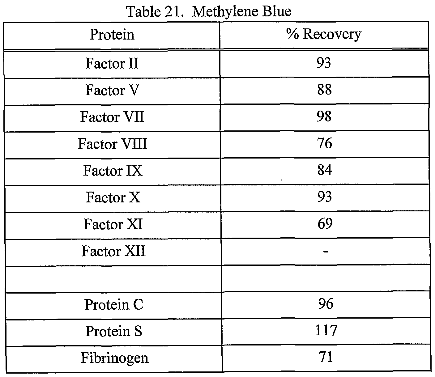


Claims
Priority Applications (4)
| Application Number | Priority Date | Filing Date | Title |
|---|---|---|---|
| DE60124095T DE60124095T2 (en) | 2000-06-15 | 2001-06-08 | METHOD FOR INACTIVATING MICROORGANISMS USING PHOTOSENSIBILIZERS |
| EP01944414A EP1289991B1 (en) | 2000-06-15 | 2001-06-08 | Method for inactivation of microorganisms using photosensitizers |
| CA002381594A CA2381594C (en) | 2000-06-15 | 2001-06-08 | Method for inactivation of microorganisms using photosensitizers |
| AU2001266829A AU2001266829A1 (en) | 2000-06-15 | 2001-06-08 | Method for inactivation of microorganisms using photosensitizers |
Applications Claiming Priority (2)
| Application Number | Priority Date | Filing Date | Title |
|---|---|---|---|
| US09/596,429 US7094378B1 (en) | 2000-06-15 | 2000-06-15 | Method and apparatus for inactivation of biological contaminants using photosensitizers |
| US09/596,429 | 2000-06-15 |
Publications (1)
| Publication Number | Publication Date |
|---|---|
| WO2001096340A1 true WO2001096340A1 (en) | 2001-12-20 |
Family
ID=24387230
Family Applications (1)
| Application Number | Title | Priority Date | Filing Date |
|---|---|---|---|
| PCT/US2001/018752 WO2001096340A1 (en) | 2000-06-15 | 2001-06-08 | Method for inactivation of microorganisms using photosensitizers |
Country Status (7)
| Country | Link |
|---|---|
| US (1) | US7094378B1 (en) |
| EP (1) | EP1289991B1 (en) |
| AT (1) | ATE343579T1 (en) |
| AU (1) | AU2001266829A1 (en) |
| CA (1) | CA2381594C (en) |
| DE (1) | DE60124095T2 (en) |
| WO (1) | WO2001096340A1 (en) |
Cited By (19)
| Publication number | Priority date | Publication date | Assignee | Title |
|---|---|---|---|---|
| WO2002043485A1 (en) * | 2000-11-28 | 2002-06-06 | Gambro, Inc. | Storage solution containing photosensitizer for inactivation of biological contaminants |
| WO2003057253A1 (en) * | 2001-12-28 | 2003-07-17 | Gambro, Inc. | Oxygen-enhanced pathogen inactivation |
| WO2003063915A1 (en) * | 2002-02-01 | 2003-08-07 | Gambro, Inc. | Reduction of contaminants in blood and blood products using photosensitizers and peak wavelengths of light |
| WO2003063902A2 (en) * | 2002-02-01 | 2003-08-07 | Gambro, Inc. | Inactivation of west nile virus and plasmodium falciparum using alloxazine-derivating photosensitizers |
| WO2003066109A1 (en) * | 2002-02-08 | 2003-08-14 | Gambro, Inc. | Nitric oxide in a pathogen inactivation process |
| WO2003090793A1 (en) * | 2002-04-24 | 2003-11-06 | Gambro, Inc. | Removal of adenine during a process of pathogen reducing blood and blood components |
| WO2003094979A1 (en) * | 2002-05-06 | 2003-11-20 | Gambro, Inc. | Method for preventing damage to or rejuvenating a cellular blood component using mitochondrial enhancer |
| EP1379640A1 (en) * | 2001-03-13 | 2004-01-14 | Triton Thalassic Technologies, Inc. | Monochromatic fluid treatment systems |
| WO2005089816A1 (en) * | 2004-03-15 | 2005-09-29 | Navigant Biotechnologies, Inc. | Methods for uniformly treating biological samples with electromagnetic radiation |
| WO2007067482A2 (en) * | 2005-12-06 | 2007-06-14 | Navigant Biotechnologies, Llc | Method for treatment and storage of blood and blood products using endogenous alloxazines and acetate |
| EP1926501A1 (en) * | 2005-09-21 | 2008-06-04 | Arecor Limited | Method for stabilisation of a protein solution by addition of hydroxyl radical quenchers and its sterilisation by ionising radiation |
| WO2009026073A1 (en) * | 2007-08-22 | 2009-02-26 | Caridianbct Biotechnologies, Llc | Prevention of transfusion related acute lung injury using riboflavin and light |
| WO2009018309A3 (en) * | 2007-08-01 | 2010-06-17 | Caridianbct Biotechnologies, Llc | Pathogen inactivation of whole blood |
| US8164073B2 (en) | 2005-12-23 | 2012-04-24 | Harald Mohr | Method for the inactivation of pathogens in donor blood, blood plasma or erythrocyte concentrates in flexible containers under agitation |
| US8173066B2 (en) | 2005-12-23 | 2012-05-08 | Forschungsgemeinschaft Der Drk Blutspendedienste E.V. | Method for irradiating thrombocyte concentrates in flexible containers with ultra-violet light |
| WO2012123412A1 (en) * | 2011-03-11 | 2012-09-20 | Deutsches Rheuma-Forschungszentrum Berlin | Flow cytometer disinfection module |
| US8778263B2 (en) | 2007-06-22 | 2014-07-15 | Maco Pharma S.A. | Irradiation apparatus for inactivating pathogens and/or leukocytes in a biological fluid and process |
| US9044523B2 (en) | 2000-06-15 | 2015-06-02 | Terumo Bct, Inc. | Reduction of contaminants in blood and blood products using photosensitizers and peak wavelengths of light |
| US10058646B2 (en) | 2006-09-19 | 2018-08-28 | Maco Pharma S.A. | Blood bag system and process for the inactivation of pathogens in platelet concentrates by use of the blood bag system |
Families Citing this family (22)
| Publication number | Priority date | Publication date | Assignee | Title |
|---|---|---|---|---|
| US7186543B1 (en) * | 1998-07-21 | 2007-03-06 | Gambro Inc. | Preparation of vaccines using a photosensitizer and light |
| US7498156B2 (en) * | 1998-07-21 | 2009-03-03 | Caridianbct Biotechnologies, Llc | Use of visible light at wavelengths of 500 to 550 nm to reduce the number of pathogens in blood and blood components |
| US8980568B2 (en) | 2001-10-11 | 2015-03-17 | Aviva Biosciences Corporation | Methods and compositions for detecting non-hematopoietic cells from a blood sample |
| US8986944B2 (en) * | 2001-10-11 | 2015-03-24 | Aviva Biosciences Corporation | Methods and compositions for separating rare cells from fluid samples |
| US20070102858A1 (en) * | 2005-11-07 | 2007-05-10 | Navigant Biotechnologies, Inc. | Clamps and methods for displacing fluid from portions of fluid containers |
| CN102318619A (en) * | 2006-01-27 | 2012-01-18 | 科安比司特生物技术有限责任公司 | Produce the method and composition of high concentration alloxazine solutions |
| WO2008008515A2 (en) | 2006-07-14 | 2008-01-17 | Aviva Biosciences Corporation | Methods and compositions for detecting rare cells from a biological sample |
| US20080234670A1 (en) * | 2007-03-15 | 2008-09-25 | Rogers C Brian | System and apparatus providing a controlled light source for medicinal applications |
| LT5567B (en) | 2008-08-06 | 2009-04-27 | Vilniaus Universitetas | Process for dezinfecting food and surfaces interfacing therewith |
| US9005530B2 (en) * | 2009-07-09 | 2015-04-14 | Donald Nevin | Compressor with an internal disinfecting UV light source |
| US9694128B1 (en) * | 2010-02-24 | 2017-07-04 | Brian LeBerthon | Device and method for administering an anti-cancer substance |
| US20140083628A1 (en) | 2012-09-27 | 2014-03-27 | Velico Medical, Inc. | Spray drier assembly for automated spray drying |
| WO2013106605A1 (en) | 2012-01-11 | 2013-07-18 | Terumo Bct Biotechnologies, Llc | Slidable clamp for port isolation |
| ES2937663T3 (en) | 2012-07-26 | 2023-03-30 | Donna J Brezinski | portable phototherapy device |
| CA2900038C (en) * | 2013-02-07 | 2021-02-09 | Rocomp Global, Llc | Electromagnetic radiation targeting devices, assemblies, systems and methods |
| US9227005B2 (en) | 2013-03-14 | 2016-01-05 | Brian J LeBerthon | Method and device for treating cancer |
| US9561184B2 (en) | 2014-09-19 | 2017-02-07 | Velico Medical, Inc. | Methods and systems for multi-stage drying of plasma |
| IT202000016864A1 (en) * | 2020-07-10 | 2022-01-10 | Emoled S R L | METHOD FOR THE INACTIVATION OF A PATHOGEN COLONY IN SPACES, SURFACES OR OBJECTS USING ELECTROMAGNETIC RADIATION |
| EP3939618A1 (en) | 2020-07-10 | 2022-01-19 | Emoled Srl | Method for inactivation of a pathogen colony in spaces, surfaces and objects using electromagnetic radiation |
| CN113414049B (en) * | 2021-06-04 | 2023-04-18 | 圣托马斯先进材料公司 | Intelligent atomizer and using method thereof |
| CN114225067B (en) * | 2021-12-22 | 2024-01-26 | 中国医学科学院输血研究所 | Blood pathogen inactivation method |
| US11841189B1 (en) | 2022-09-15 | 2023-12-12 | Velico Medical, Inc. | Disposable for a spray drying system |
Citations (2)
| Publication number | Priority date | Publication date | Assignee | Title |
|---|---|---|---|---|
| US5360734A (en) * | 1991-06-21 | 1994-11-01 | Baxter International, Inc. | Method for inactivating pathogens in erythrocytes using photoactive compounds and plasma protein reduction |
| WO2000004930A2 (en) * | 1998-07-21 | 2000-02-03 | Gambro, Inc. | Method for inactivation of microorganisms using photosensitizers |
Family Cites Families (102)
| Publication number | Priority date | Publication date | Assignee | Title |
|---|---|---|---|---|
| US683690A (en) | 1900-06-13 | 1901-10-01 | Charles M Johnson | Apparatus for sterilizing disease-germs. |
| US1733239A (en) | 1929-01-31 | 1929-10-29 | Donald E Roberts | Applicator for conducting ultra-violet rays |
| US1961700A (en) | 1932-07-29 | 1934-06-05 | Gen Electric Vapor Lamp Co | Apparatus for sterilizing articles by ultraviolet radiation |
| US2056614A (en) | 1933-06-13 | 1936-10-06 | Gen Electric Vapor Lamp Co | Ultraviolet sterilizer |
| US2111491A (en) | 1935-01-26 | 1938-03-15 | Winthrop Chem Co Inc | Process of preparing phosphoric acid esters of hydroxyalkyl isoalloxazines |
| US2212330A (en) | 1938-05-03 | 1940-08-20 | Albert G Thomas | Sterilizing device |
| US2212230A (en) | 1938-05-28 | 1940-08-20 | Internat Telephone Dev Co Inc | Airplane guiding beacon |
| US2340890A (en) | 1941-02-25 | 1944-02-08 | Lang Alphonse | Method and apparatus for sterilizing, preserving, and irradiating of various liquid substances |
| US2340980A (en) | 1942-08-14 | 1944-02-08 | Mexican American Hat Company | Sweatband structure |
| US2654735A (en) | 1949-07-29 | 1953-10-06 | Us Vitamin Corp | Process for the production of derivatives of 9-polyhydroxyalkylisoalloxazines and products obtained |
| US2825729A (en) | 1955-05-24 | 1958-03-04 | Upjohn Co | Isoalloxazines |
| US3189598A (en) | 1960-01-14 | 1965-06-15 | Yagi Kunio | Fatty acid esters of riboflavin |
| US3456053A (en) | 1966-05-06 | 1969-07-15 | Pfizer & Co C | Inactivated hog cholera virus vaccine |
| US3683183A (en) | 1969-06-04 | 1972-08-08 | Radiation Machinery Corp | A flow-through irradiator for the extra corporeal irradiation of fluid |
| US3705985A (en) | 1969-06-27 | 1972-12-12 | Nuclear Associates Inc | Fluid irradiator and process for its manufacture |
| US3683177A (en) | 1970-06-30 | 1972-08-08 | Louis P Veloz | Sterilization of a fluid by ultraviolet radiation |
| US3852032A (en) | 1971-06-07 | 1974-12-03 | Uroptics Int Inc | Process for sterilizing hydrophilic gelatin lenses having ultraviolet stabilizers |
| US3776694A (en) | 1972-04-04 | 1973-12-04 | L Leittl | Germicidal toiletry cabinet for different personal hygiene items |
| US3926556A (en) | 1973-05-30 | 1975-12-16 | Raymond Marcel Gut Boucher | Biocidal electromagnetic synergistic process |
| US3864081A (en) | 1973-06-12 | 1975-02-04 | Spectroderm International Inc | Apparatus for sterilizing biologic material and the like by ultra-violet irradiation |
| US3920650A (en) | 1973-09-19 | 1975-11-18 | Morton Norwich Products Inc | Isoalloxazines |
| US3894236A (en) | 1973-12-10 | 1975-07-08 | Wayne K Hazelrigg | Device for irradiating fluids |
| US3927325A (en) | 1974-07-10 | 1975-12-16 | Us Energy | Tissue irradiator |
| US4181128A (en) | 1975-11-28 | 1980-01-01 | Massachusetts Institute Of Technology | Virus inactivation applicator and the like |
| US4139348A (en) | 1975-11-28 | 1979-02-13 | Massachusetts Institute Of Technology | Electrochemical process and apparatus to control the chemical state of a material |
| US4305390A (en) | 1975-11-28 | 1981-12-15 | Massachusetts Institute Of Technology | Method for generating oxygen in an excited electronic state and inactivation of microorganisms |
| US4173631A (en) | 1976-08-23 | 1979-11-06 | Merck & Co., Inc. | 7-Methyl-8-methylamino-10-(1'-D-ribityl)isoalloxazine |
| US4196281A (en) | 1976-10-20 | 1980-04-01 | Regents Of The University Of California | Psoralens |
| US4124598A (en) | 1976-10-20 | 1978-11-07 | Hoffmann-La Roche Inc. | Psoralens |
| US4169204A (en) | 1976-10-20 | 1979-09-25 | Regents Of The University Of California | Psoralens |
| US4424201A (en) | 1978-11-28 | 1984-01-03 | Rockefeller University | Employment of a mereyanine dye for the detection of malignant leukocytic cells |
| IT1166343B (en) | 1979-08-20 | 1987-04-29 | Francarosa Baccichetti | FUROCUMARINE FOR THE PHOTOCHEMOTHERAPY OF FSORIASIS AND OTHER SENSITIVE SKIN DISEASES |
| US4321918A (en) | 1979-10-23 | 1982-03-30 | Clark Ii William T | Process for suppressing immunity to transplants |
| US4398906A (en) | 1979-12-11 | 1983-08-16 | Frederic A. Bourke, Jr. | Method for externally treating the blood |
| US4428744A (en) | 1979-12-11 | 1984-01-31 | Frederic A. Bourke, Jr. | Method and system for externally treating the blood |
| US4321919A (en) | 1979-12-11 | 1982-03-30 | Leukocyte Research, Inc. | Method and system for externally treating human blood |
| DE8007265U1 (en) | 1980-03-17 | 1981-08-27 | ESPE Fabrik pharmazeutischer Präparate GmbH, 8031 Seefeld | DEVICE FOR TREATING SPARE PARTS |
| US4336809A (en) | 1980-03-17 | 1982-06-29 | Burleigh Instruments, Inc. | Human and animal tissue photoradiation system and method |
| US4481167A (en) | 1980-04-11 | 1984-11-06 | The Dow Chemical Company | Sanitizing complexes of polyoxazolines or polyoxazines and polyhalide anions |
| US4398031A (en) | 1980-06-11 | 1983-08-09 | The Regents Of The University Of California | Coumarin derivatives and method for synthesizing 5'-methyl psoralens therefrom |
| JPS616899Y2 (en) | 1981-04-27 | 1986-03-03 | ||
| US4464166A (en) | 1981-06-12 | 1984-08-07 | Frederic A. Bourke, Jr. | Method for externally treating the blood |
| US4612007A (en) | 1981-06-16 | 1986-09-16 | Edelson Richard Leslie | Method and system for externally treating the blood |
| WO1983000811A1 (en) | 1981-09-08 | 1983-03-17 | Stancon, Alexei | Photoradiation method and arrangement |
| US4467206A (en) | 1981-12-14 | 1984-08-21 | Extracorporeal Medical Specialties, Inc. | Method and apparatus for the irradiation of fluids |
| US4456512A (en) | 1982-03-10 | 1984-06-26 | The Dow Chemical Company | Photochemical reactor and method |
| US4649151A (en) | 1982-09-27 | 1987-03-10 | Health Research, Inc. | Drugs comprising porphyrins |
| CA1216518A (en) | 1982-11-01 | 1987-01-13 | Gail A. Rock | Plasma-free medium for platelet storage |
| US4613322A (en) | 1982-12-08 | 1986-09-23 | Edelson Richard Leslie | Method and system for externally treating the blood |
| US4684521A (en) | 1982-12-08 | 1987-08-04 | Frederic A. Bourke, Jr. | Method and system for externally treating the blood |
| US4683889A (en) | 1983-03-29 | 1987-08-04 | Frederic A. Bourke, Jr. | Method and system for externally treating the blood |
| US4748120A (en) | 1983-05-02 | 1988-05-31 | Diamond Scientific Co. | Photochemical decontamination treatment of whole blood or blood components |
| US4727027A (en) | 1983-05-02 | 1988-02-23 | Diamond Scientific Co. | Photochemical decontamination treatment of whole blood or blood components |
| US4946438A (en) | 1983-09-01 | 1990-08-07 | The Trustees Of Columbia University In The City Of New York | Process for development of acceptance of transplanted organs and tissues |
| US4861704A (en) | 1983-09-01 | 1989-08-29 | The Trustees Of Columbia University In The City Of New York | Processes for development of acceptance of transplanted organs and tissues |
| US4992363A (en) | 1983-11-09 | 1991-02-12 | Thomas Jefferson University | Method for preparing glucose free media for storing blood platelets |
| US4693981A (en) | 1983-12-20 | 1987-09-15 | Advanced Genetics Research Institute | Preparation of inactivated viral vaccines |
| US4604356A (en) | 1983-12-21 | 1986-08-05 | Miles Laboratories, Inc. | Purification of flavin adenine dinucleotide synthetase |
| CH657864A5 (en) | 1984-02-17 | 1986-09-30 | Ciba Geigy Ag | WATER-SOLUBLE PHTHALOCYANINE COMPOUNDS AND THE USE THEREOF AS PHOTOACTIVATORS. |
| US4493981A (en) | 1984-03-05 | 1985-01-15 | General Electric Company | Boil dry protection system for cooking appliance |
| JPH0622222B2 (en) | 1984-09-18 | 1994-03-23 | 株式会社東芝 | Light processing equipment |
| JPS6176160A (en) | 1984-09-21 | 1986-04-18 | 松永 是 | Cell killing method |
| US4576143A (en) | 1984-10-05 | 1986-03-18 | Clark Iii William T | Method of immune modification by means of extracorporeal irradiation of the blood |
| US4573962A (en) | 1984-10-29 | 1986-03-04 | Extracorporeal Medical Specialties, Inc. | Cassette drawer assembly for photoactivation patient treatment system |
| US4573960A (en) | 1984-10-29 | 1986-03-04 | Extracorporeal Medical Specialties, Inc. | Three phase irradiation treatment process |
| US4737140A (en) | 1984-10-29 | 1988-04-12 | Mcneilab, Inc. | Irradiation chamber for photoactivation patient treatment system |
| US4708715A (en) | 1984-10-29 | 1987-11-24 | Mcneilab, Inc. | Light array assembly for photoactivation patient treatment system |
| US4573961A (en) | 1984-10-29 | 1986-03-04 | Extracorporeal Medical Specialties, Inc. | Electronic control methods for puvapheresis apparatus |
| US4568328A (en) | 1984-10-29 | 1986-02-04 | Extracorporeal Medical Specialties, Inc. | Automated photophoresis blood portion control methods and apparatus |
| US4623328A (en) | 1984-10-29 | 1986-11-18 | Mcneilab, Inc. | Pump monitor for photoactivation patient treatment system |
| US4578056A (en) | 1984-10-29 | 1986-03-25 | Extracorporeal Medical Specialties, Inc. | Patient photopheresis treatment apparatus and method |
| US4596547A (en) | 1984-10-29 | 1986-06-24 | Mcneilab, Inc. | Valve apparatus for photoactivation patient treatment system |
| US4608255A (en) | 1985-01-31 | 1986-08-26 | American National Red Cross | Biocompatible method for in situ production of functional platelets and product produced thereby lacking immunogenicity |
| DD249143A3 (en) | 1985-03-20 | 1987-09-02 | Ilmenau Tech Hochschule | DEVICE FOR THE PHYSIOLOGICAL THERAPEUTICALLY ACTIVE OPTICAL IRRADIATION OF OVERLAPPED VENEER BLOOD |
| US4683195A (en) | 1986-01-30 | 1987-07-28 | Cetus Corporation | Process for amplifying, detecting, and/or-cloning nucleic acid sequences |
| US4651739A (en) | 1985-04-08 | 1987-03-24 | The General Hospital Corporation | Light-induced killing of carcinoma cells |
| US4998931A (en) | 1985-07-05 | 1991-03-12 | Puget Sound Blood Center | Method of reducing immunogenicity and inducing immunologic tolerance |
| US4930516B1 (en) | 1985-11-13 | 1998-08-04 | Laser Diagnostic Instr Inc | Method for detecting cancerous tissue using visible native luminescence |
| US4695460A (en) | 1986-03-19 | 1987-09-22 | American Red Cross | Synthetic, plasma-free, transfusible platelet storage medium |
| US4961928A (en) | 1986-03-19 | 1990-10-09 | American Red Cross | Synthetic, plasma-free, transfusible storage medium for red blood cells and platelets |
| US4726949A (en) | 1986-08-26 | 1988-02-23 | Baxter Travenol Laboratories, Inc. | Irradiation of blood products |
| US4952812A (en) | 1986-08-26 | 1990-08-28 | Baxter International Inc. | Irradiation of blood products |
| US4866282A (en) | 1986-08-26 | 1989-09-12 | Baxter International Inc. | Irradiation of blood products |
| US4775625A (en) | 1986-11-21 | 1988-10-04 | The Medical College Of Wisconsin, Inc. | Inactivating enveloped viruses with a merocyanine dye |
| US4915683A (en) | 1986-11-21 | 1990-04-10 | The Medical College Of Wisconsin, Inc. | Antiviral method, agents and apparatus |
| US4878891A (en) | 1987-06-25 | 1989-11-07 | Baylor Research Foundation | Method for eradicating infectious biological contaminants in body tissues |
| US4833165A (en) | 1987-10-07 | 1989-05-23 | Louderback Allan Lee | Method of inactivating HTLV-III virus in blood |
| US4880788A (en) | 1987-10-30 | 1989-11-14 | Baylor College Of Medicine | Method for preventing and treating thrombosis |
| DE3805459A1 (en) | 1988-02-22 | 1989-08-31 | Biotest Pharma Gmbh | METHOD FOR STERILIZING BLOOD, PLASMA, BLOOD AND PLASMA DERIVATIVES, CELL SUSPENSIONS OR THE LIKE |
| DE3824647A1 (en) | 1988-07-20 | 1990-02-01 | Wedeco Entkeimungsanlagen | DEVICE FOR IRRADIATING MEDIA BY UV LIGHT |
| EP0386241A4 (en) | 1988-08-23 | 1990-10-03 | Radiotech Inst An | Light-guiding device for medical treatment |
| US4994367A (en) | 1988-10-07 | 1991-02-19 | East Carolina University | Extended shelf life platelet preparations and process for preparing the same |
| US4950665A (en) | 1988-10-28 | 1990-08-21 | Oklahoma Medical Research Foundation | Phototherapy using methylene blue |
| US4960408A (en) | 1989-01-10 | 1990-10-02 | Klainer Albert S | Treatment methods and vaccines for stimulating an immunological response against retroviruses |
| US4921473A (en) | 1989-02-02 | 1990-05-01 | Therakos, Inc. | Multicomponent fluid separation and irradiation system |
| US4978688A (en) | 1989-03-24 | 1990-12-18 | Louderback Allan Lee | Method of treating white blood cells |
| US4999375A (en) | 1989-04-11 | 1991-03-12 | Hoffmann-La Roche Inc. | Psoralen reagent compositions for extracorporeal treatment of blood |
| US6187572B1 (en) * | 1990-04-16 | 2001-02-13 | Baxter International Inc. | Method of inactivation of viral and bacterial blood contaminants |
| US6235508B1 (en) * | 1995-06-07 | 2001-05-22 | Baxter International Inc. | Method of inactivation of viral and bacterial blood contaminants |
| US6277337B1 (en) * | 1998-07-21 | 2001-08-21 | Gambro, Inc. | Method and apparatus for inactivation of biological contaminants using photosensitizers |
| US6268120B1 (en) * | 1999-10-19 | 2001-07-31 | Gambro, Inc. | Isoalloxazine derivatives to neutralize biological contaminants |
| US6548241B1 (en) * | 2000-11-28 | 2003-04-15 | Gambro, Inc. | Storage solution containing photosensitizer for inactivation of biological contaminants |
-
2000
- 2000-06-15 US US09/596,429 patent/US7094378B1/en not_active Expired - Fee Related
-
2001
- 2001-06-08 EP EP01944414A patent/EP1289991B1/en not_active Expired - Lifetime
- 2001-06-08 DE DE60124095T patent/DE60124095T2/en not_active Expired - Lifetime
- 2001-06-08 AT AT01944414T patent/ATE343579T1/en not_active IP Right Cessation
- 2001-06-08 CA CA002381594A patent/CA2381594C/en not_active Expired - Lifetime
- 2001-06-08 AU AU2001266829A patent/AU2001266829A1/en not_active Abandoned
- 2001-06-08 WO PCT/US2001/018752 patent/WO2001096340A1/en active IP Right Grant
Patent Citations (2)
| Publication number | Priority date | Publication date | Assignee | Title |
|---|---|---|---|---|
| US5360734A (en) * | 1991-06-21 | 1994-11-01 | Baxter International, Inc. | Method for inactivating pathogens in erythrocytes using photoactive compounds and plasma protein reduction |
| WO2000004930A2 (en) * | 1998-07-21 | 2000-02-03 | Gambro, Inc. | Method for inactivation of microorganisms using photosensitizers |
Cited By (37)
| Publication number | Priority date | Publication date | Assignee | Title |
|---|---|---|---|---|
| US9044523B2 (en) | 2000-06-15 | 2015-06-02 | Terumo Bct, Inc. | Reduction of contaminants in blood and blood products using photosensitizers and peak wavelengths of light |
| WO2002043485A1 (en) * | 2000-11-28 | 2002-06-06 | Gambro, Inc. | Storage solution containing photosensitizer for inactivation of biological contaminants |
| EP1379640A4 (en) * | 2001-03-13 | 2006-09-27 | Triton Thalassic Technologies | Monochromatic fluid treatment systems |
| EP1379640A1 (en) * | 2001-03-13 | 2004-01-14 | Triton Thalassic Technologies, Inc. | Monochromatic fluid treatment systems |
| WO2003057253A1 (en) * | 2001-12-28 | 2003-07-17 | Gambro, Inc. | Oxygen-enhanced pathogen inactivation |
| JP4704684B2 (en) * | 2002-02-01 | 2011-06-15 | カリディアンビーシーティ バイオテクノロジーズ,エルエルシー | Reduction of contamination in blood and blood products using photosensitizers and peak wavelengths of light |
| WO2003063902A3 (en) * | 2002-02-01 | 2003-09-04 | Gambro Inc | Inactivation of west nile virus and plasmodium falciparum using alloxazine-derivating photosensitizers |
| JP2005516978A (en) * | 2002-02-01 | 2005-06-09 | ガンブロ インコーポレーテッド | Reduction of contamination in blood and blood products using photosensitizers and peak wavelengths of light |
| JP2005519920A (en) * | 2002-02-01 | 2005-07-07 | ガンブロ インコーポレーテッド | Inactivation of West Nile virus and malaria using photosensitizers |
| WO2003063915A1 (en) * | 2002-02-01 | 2003-08-07 | Gambro, Inc. | Reduction of contaminants in blood and blood products using photosensitizers and peak wavelengths of light |
| WO2003063902A2 (en) * | 2002-02-01 | 2003-08-07 | Gambro, Inc. | Inactivation of west nile virus and plasmodium falciparum using alloxazine-derivating photosensitizers |
| WO2003066109A1 (en) * | 2002-02-08 | 2003-08-14 | Gambro, Inc. | Nitric oxide in a pathogen inactivation process |
| WO2003090793A1 (en) * | 2002-04-24 | 2003-11-06 | Gambro, Inc. | Removal of adenine during a process of pathogen reducing blood and blood components |
| US7183045B2 (en) | 2002-04-24 | 2007-02-27 | Gambro Inc. | Removal of adenine during a pathogen reduction process in whole blood or red blood cell by dilution |
| US8679736B2 (en) | 2002-04-24 | 2014-03-25 | Terumo Bct Biotechnologies, Llc | Removal of adenine during a pathogen reduction process in whole blood or red blood cells by dilution |
| WO2003094979A1 (en) * | 2002-05-06 | 2003-11-20 | Gambro, Inc. | Method for preventing damage to or rejuvenating a cellular blood component using mitochondrial enhancer |
| KR101184153B1 (en) * | 2004-03-15 | 2012-09-18 | 테루모 비씨티 바이오테크놀로지스, 엘엘씨 | Methods for uniformly treating biological samples with electromagnetic radiation |
| JP2007531569A (en) * | 2004-03-15 | 2007-11-08 | ナビガント・バイオテクノロジーズ、インコーポレイテッド | Uniform treatment of biological samples by electromagnetic radiation |
| WO2005089816A1 (en) * | 2004-03-15 | 2005-09-29 | Navigant Biotechnologies, Inc. | Methods for uniformly treating biological samples with electromagnetic radiation |
| US8296071B2 (en) | 2004-03-15 | 2012-10-23 | Terumo Bct Biotechnologies, Llc | Methods for uniformly treating biological samples with electromagnetic radiation |
| CN102188728B (en) * | 2004-03-15 | 2012-05-23 | 科安比司特生物技术有限责任公司 | Methods for uniformly treating biological samples with electromagnetic radiation |
| AU2010241297B2 (en) * | 2004-03-15 | 2012-05-17 | Terumo Bct Biotechnologies, Llc | Methods for uniformly treating biological samples with electromagnetic radiation |
| JP2011172940A (en) * | 2004-03-15 | 2011-09-08 | Caridianbct Biotechnologies Llc | Method for uniformly treating biological sample with electromagnetic radiation |
| EP1926501A1 (en) * | 2005-09-21 | 2008-06-04 | Arecor Limited | Method for stabilisation of a protein solution by addition of hydroxyl radical quenchers and its sterilisation by ionising radiation |
| WO2007067482A3 (en) * | 2005-12-06 | 2009-02-26 | Navigant Biotechnologies Llc | Method for treatment and storage of blood and blood products using endogenous alloxazines and acetate |
| WO2007067482A2 (en) * | 2005-12-06 | 2007-06-14 | Navigant Biotechnologies, Llc | Method for treatment and storage of blood and blood products using endogenous alloxazines and acetate |
| US8173066B2 (en) | 2005-12-23 | 2012-05-08 | Forschungsgemeinschaft Der Drk Blutspendedienste E.V. | Method for irradiating thrombocyte concentrates in flexible containers with ultra-violet light |
| US8164073B2 (en) | 2005-12-23 | 2012-04-24 | Harald Mohr | Method for the inactivation of pathogens in donor blood, blood plasma or erythrocyte concentrates in flexible containers under agitation |
| US8525128B2 (en) | 2005-12-23 | 2013-09-03 | Blutspendedienst Der Landesverbande Des Deutschen Roten Kreuzes Niedersachsen, Sachsen-Anhalt, Thuringen, Oldenburg Und Bremen G.G.M.B.H. | Method for the inactivation of pathogens in donor blood, blood plasma or erythrocyte concentrates in flexible containers under agitation |
| US10058646B2 (en) | 2006-09-19 | 2018-08-28 | Maco Pharma S.A. | Blood bag system and process for the inactivation of pathogens in platelet concentrates by use of the blood bag system |
| US8778263B2 (en) | 2007-06-22 | 2014-07-15 | Maco Pharma S.A. | Irradiation apparatus for inactivating pathogens and/or leukocytes in a biological fluid and process |
| US9320817B2 (en) | 2007-06-22 | 2016-04-26 | Maco Pharma S.A. | Irradiation apparatus for inactivating pathogens and/or leukocytes in a biological fluid and process |
| JP2010535235A (en) * | 2007-08-01 | 2010-11-18 | カリディアンビーシーティ バイオテクノロジーズ,エルエルシー | Pathogenic inactivation of whole blood |
| WO2009018309A3 (en) * | 2007-08-01 | 2010-06-17 | Caridianbct Biotechnologies, Llc | Pathogen inactivation of whole blood |
| WO2009026073A1 (en) * | 2007-08-22 | 2009-02-26 | Caridianbct Biotechnologies, Llc | Prevention of transfusion related acute lung injury using riboflavin and light |
| WO2012123412A1 (en) * | 2011-03-11 | 2012-09-20 | Deutsches Rheuma-Forschungszentrum Berlin | Flow cytometer disinfection module |
| US9498550B2 (en) | 2011-03-11 | 2016-11-22 | Deutsches Rheuma-Forschungszentrum Berlin | Flow cytometer disinfection module |
Also Published As
| Publication number | Publication date |
|---|---|
| AU2001266829A1 (en) | 2001-12-24 |
| EP1289991A1 (en) | 2003-03-12 |
| DE60124095T2 (en) | 2007-06-06 |
| CA2381594A1 (en) | 2001-12-20 |
| DE60124095D1 (en) | 2006-12-07 |
| CA2381594C (en) | 2009-03-24 |
| EP1289991B1 (en) | 2006-10-25 |
| US7094378B1 (en) | 2006-08-22 |
| ATE343579T1 (en) | 2006-11-15 |
Similar Documents
| Publication | Publication Date | Title |
|---|---|---|
| US7094378B1 (en) | Method and apparatus for inactivation of biological contaminants using photosensitizers | |
| US6277337B1 (en) | Method and apparatus for inactivation of biological contaminants using photosensitizers | |
| EP1047458B1 (en) | Method for inactivation of microorganisms using photosensitizers | |
| CA2397862C (en) | Storage solution containing photosensitizer for inactivation of biological contaminants | |
| JP2004500316A5 (en) | ||
| EP1286996A1 (en) | Method and apparatus for inactivation of biological contaminants using photosensitizers | |
| US20030215784A1 (en) | Method and apparatus for inactivation of biological contaminants using photosensitizers | |
| AU770614B2 (en) | Method and apparatus for inactivation of biological contaminants using photosensitizers | |
| CA2585179C (en) | Method and apparatus for inactivation of biological contaminants using photosensitizers | |
| CZ20001406A3 (en) | Inactivation process of biological contaminants by making use of photosensitizers and apparatus for making the same | |
| MXPA00002800A (en) | Method and apparatus for inactivation of biological contaminants using photosensitizers |
Legal Events
| Date | Code | Title | Description |
|---|---|---|---|
| AK | Designated states |
Kind code of ref document: A1 Designated state(s): AE AG AL AM AT AU AZ BA BB BG BR BY BZ CA CH CN CR CU CZ DE DK DM DZ EE ES FI GB GD GE GH GM HR HU ID IL IN IS JP KE KG KP KR KZ LC LK LR LS LT LU LV MA MD MG MK MN MW MX MZ NO NZ PL PT RO RU SD SE SG SI SK SL TJ TM TR TT TZ UA UG UZ VN YU ZA ZW |
|
| AL | Designated countries for regional patents |
Kind code of ref document: A1 Designated state(s): GH GM KE LS MW MZ SD SL SZ TZ UG ZW AM AZ BY KG KZ MD RU TJ TM AT BE CH CY DE DK ES FI FR GB GR IE IT LU MC NL PT SE TR BF BJ CF CG CI CM GA GN GW ML MR NE SN TD TG |
|
| WWE | Wipo information: entry into national phase |
Ref document number: 2381594 Country of ref document: CA |
|
| 121 | Ep: the epo has been informed by wipo that ep was designated in this application | ||
| REG | Reference to national code |
Ref country code: DE Ref legal event code: 8642 |
|
| WWE | Wipo information: entry into national phase |
Ref document number: 2001944414 Country of ref document: EP |
|
| WWP | Wipo information: published in national office |
Ref document number: 2001944414 Country of ref document: EP |
|
| NENP | Non-entry into the national phase |
Ref country code: JP |
|
| WWG | Wipo information: grant in national office |
Ref document number: 2001944414 Country of ref document: EP |